Esthetic crown lengthening is a periodontal-restorative procedure that increases visible crown height and refines gingival contours in the esthetic zone to achieve predictable anterior esthetics. Unlike functional crown lengthening, which exposes sound tooth structure for retention and resistance form, the esthetic version is driven entirely by the final restoration margin or the desired smile design. The core planning rule is simple but non-negotiable: plan to the final restorative margin, not to the current gingival level, and maintain approximately 3 mm from that intended margin to the alveolar crest to preserve supracrestal tissue attachment.
Immediate clinical takeaways:
- A “gummy smile” is a sign, not a diagnosis. Correctly identifying the etiology is essential before planning any surgical intervention.
- The supracrestal tissue attachment (formerly “biologic width”) averages ~2.04 mm, and adding mean sulcus depth (~0.69 mm) gives the practical 3 mm surgical landmark.
- Staging and provisional management reduce unesthetic sequelae and margin unpredictability in high-demand esthetic cases.
Pro Tip: Never design your incision around where the gingival margin sits today. Fabricate a provisional restoration or wax-up first, confirm the final margin position, and let that drive every surgical decision.
What are the right indications for esthetic crown lengthening?
Indications and differential diagnosis
The most common indication is excessive gingival display, but the cause of that display determines whether surgery is appropriate at all. A gummy smile is a sign, not a diagnosis, and misidentifying the etiology leads to the wrong treatment.
Primary indications for esthetic crown lengthening:
- Altered passive eruption (APE): alveolar crest within 2 mm of the CEJ, with excess gingival tissue covering anatomic crown height
- Short clinical crowns from tooth wear, attrition, or incomplete eruption
- Uneven gingival margins requiring harmonization before veneer or crown placement
- Restorative margin needs where the planned margin requires osseous recontouring for adequate supracrestal tissue attachment
Differential diagnosis for excessive gingival display:
- Altered passive eruption: excess gingival tissue with normal or near-normal crown length; CEJ is subgingival or at the crest
- Gingival enlargement: drug-induced (cyclosporine, calcium channel blockers, phenytoin), inflammatory, or hereditary; tissue is hyperplastic, not just coronally positioned
- Short clinical crowns from wear: CEJ is in normal position; crown length is reduced by attrition or erosion
- Hypermobile upper lip: normal gingival levels at rest; excessive display only at full smile; lip dynamics are the culprit
- Vertical maxillary excess (VME): skeletal etiology; excessive display persists even with normal gingival levels; orthognathic surgery may be required
Measurement protocol before any surgical decision:
- Full-face and retracted photographs at rest and full smile
- Full-mouth periodontal charting with probing depths and recession measurements
- Bone sounding under local anesthesia to locate the CEJ and alveolar crest
- CEJ-to-bone crest measurement (the key number: ≥2 mm suggests gingivectomy may suffice; <2 mm means osseous resection is required)
- Keratinized tissue width assessment (minimum 2 mm desirable post-surgery)
- Lip dynamics: measure gingival display at rest vs. full smile
Periapical radiographs are the standard imaging tool for CEJ-to-crest assessment. CBCT adds value when root morphology, bone thickness, or APE classification is uncertain. A CBCT-based modified APE classification predicts whether flapless approaches are feasible and helps identify the roughly one-third of patients whose gingival display can be reduced to under 4 mm with a less invasive approach. Digital smile design and diagnostic wax-ups are not optional extras in esthetic cases; they define the surgical endpoint.
Contraindications and case selection flags:
- Insufficient keratinized tissue width (<2 mm) without a plan to augment
- Unfavorable crown-to-root ratio after anticipated bone removal
- Active periodontal disease or uncontrolled systemic conditions
- Skeletal etiology (VME) where surgery alone will not resolve the display
- Hypermobile lip as the sole cause of excessive gingival display
- Patient with unrealistic esthetic expectations or poor compliance history
| Etiology |
Key Clinical Cue |
Surgical Candidate? |
| Altered passive eruption |
CEJ subgingival; excess tissue |
Yes, primary indication |
| Gingival enlargement |
Hyperplastic tissue; drug history |
Possibly, after medical management |
| Tooth wear / short crowns |
Normal CEJ position; worn incisal edges |
Possibly, combined with restorative |
| Hypermobile upper lip |
Normal gingival levels at rest |
No; consider lip repositioning or botulinum toxin |
| Vertical maxillary excess |
Skeletal display; normal gingival levels |
No; orthognathic referral |
How does esthetic crown lengthening differ from functional crown lengthening?
The goal and the surgical reference point are different, and that changes everything about how you plan the case.
Esthetic crown lengthening uses the CEJ as the primary reference. The objective is to expose more of the anatomic crown, create a harmonious gingival line, and establish supracrestal tissue attachment around natural tooth structure. The incision follows the esthetic smile design, and the final flap position sits at or slightly coronal to the CEJ. Buccal tissue only is typically involved; the interdental papillae and palatal flap are left undisturbed.
Functional (restorative) crown lengthening uses the final restorative margin (FRM) as the reference. The goal is to expose sound tooth structure for retention and resistance form, and the alveolar crest must end up at least 3 mm apical to that margin. Buccal and palatal/lingual flaps are raised, and vertical releasing incisions are usually needed to apically position the flap adequately.
Key practical implications:
- In esthetic cases, no vertical releasing incisions are made; this preserves papillary architecture and avoids scarring in the smile zone.
- Osseous surgery in esthetic cases follows the CEJ contour; in functional cases it follows the FRM.
- Surgical stents are often needed for esthetic cases to guide incision placement; functional cases use provisional crowns as the reference.
- Final esthetic outcomes differ: esthetic crown lengthening produces a natural smile with proportional crowns; functional crown lengthening often results in square-looking crowns with long contact points.
| Factor |
Esthetic Crown Lengthening |
Functional Crown Lengthening |
| Primary goal |
Harmonious smile line; expose anatomic crown |
Expose sound tooth structure for restoration |
| Surgical reference |
CEJ |
Final restorative margin (FRM) |
| Flap design |
Buccal only; no vertical releases |
Buccal + palatal/lingual; vertical releases common |
| Osseous target |
Crest 2–3 mm below CEJ |
Crest ≥3 mm below FRM |
| Surgical stent |
Often required |
Not required (provisional crown used) |
| Healing/timing |
3–6 months for esthetic maturation |
6 weeks minimum for posterior/functional cases |
| Final result |
Natural proportional crowns |
Square crowns, long contact points |
How should you plan a restoration-driven esthetic crown lengthening case?
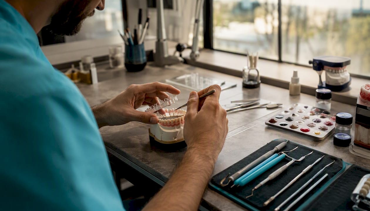
Step-by-step planning workflow
Restoration-driven planning is the single most important concept in this procedure. Bone removal is dictated by the desired restorative margin position, not by the current gingival level. Every step in the workflow flows from that principle.
- Initial records: Full-face and retracted photographs, periapical radiographs, periodontal charting, bone sounding, and a diagnostic wax-up or digital smile design.
- Define the final margin position: Work with the restorative clinician to confirm where the definitive restoration margin will sit. This is the fixed point that drives all subsequent surgical decisions.
- Fabricate a provisional restoration or surgical guide: The provisional contours define the desired gingival zenith, embrasure form, and crown proportions. A surgical guide derived from this provisional is the most reliable intraoperative reference.
- Determine osseous resection needs: Measure CEJ-to-crest distance clinically and radiographically. If the crest is within 2 mm of the CEJ (or within 3 mm of the planned margin), osseous resection is required.
- Classify the case and select the staging approach: Use the Type I/II classification (adequate vs. inadequate keratinized tissue) and subgroup A/B (CEJ-to-bone ≥2 mm vs. <2 mm) to select the correct technique. Type II/III scenarios with extensive osseous needs benefit from staged surgery.
- Interdisciplinary communication: Before surgery, the periodontist and restorative clinician must agree on final margin position, desired crown length and proportions, provisional restoration plan, and the post-surgical timeline.
- Provisional restoration strategy: Provisionals placed immediately after surgery control gingival margin position during healing. They must not impinge on the healing supracrestal tissue attachment.
Staging reduces unesthetic sequelae. When soft-tissue recontouring alone is performed first, the clinician can assess tissue response before committing to osseous resection. This is particularly valuable in cases where the extent of bone removal is uncertain, or where open embrasures and black triangles are a risk.
Interdisciplinary checklist before surgery:
- Agreed final margin position (documented with photographs and mounted models)
- Confirmed crown length and gingival zenith positions
- Provisional restoration fabricated and approved by both clinician and patient
- Surgical guide or stent derived from provisional design
- CBCT reviewed if root morphology or bone thickness is uncertain
- Informed consent completed, including recession risk, sensitivity, and timing of final restoration
Pro Tip: Fabricate the surgical guide from the approved provisional wax-up, not from the current tooth position. Intraoperatively, seat the guide to confirm incision placement before making the first cut. This one step eliminates the most common planning error: removing too little bone because the surgeon is referencing the wrong landmark.
What are the surgical technique options and when do you use each?
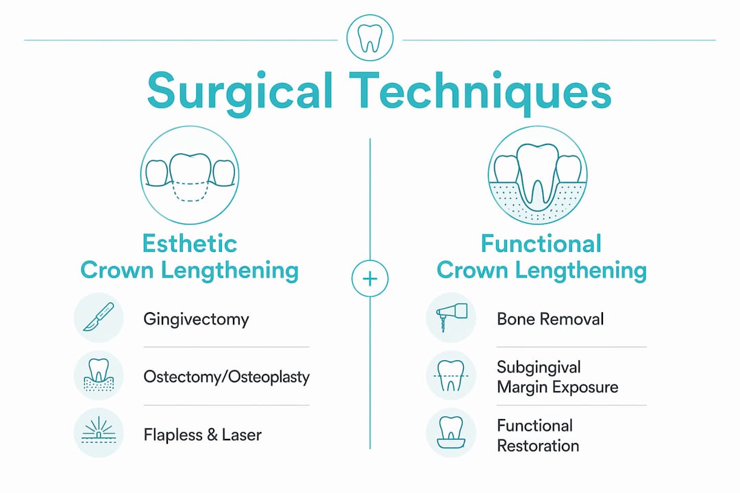
Gingivectomy and gingivoplasty
Use this when keratinized tissue is adequate (≥2 mm) and the CEJ-to-bone distance is ≥2 mm. The procedure is soft-tissue only: no flap, no bone removal. The incision follows the esthetic smile design using the CEJ as the reference. Adequate keratinized tissue width and a favorable CEJ-to-crest distance are the two prerequisites that make this the simplest, most predictable option.
Apically positioned flap with ostectomy/osteoplasty
This is the workhorse technique for most esthetic crown lengthening cases. It is required when the CEJ-to-bone distance is <2 mm, or when the planned restorative margin requires the crest to be repositioned apically. The target is ~3 mm from the desired margin to the alveolar crest after osseous resection.
Intraoperative steps:
- Administer local anesthesia; confirm bone sounding landmarks.
- Place the surgical guide or seat the provisional to confirm incision line.
- Make the initial buccal incision following the esthetic smile line, using the CEJ as the reference. No vertical releasing incisions in the esthetic zone unless absolutely necessary.
- Reflect a full-thickness buccal flap only; do not raise the interdental papillae or the palatal flap.
- Perform ostectomy and osteoplasty to position the alveolar crest ~3 mm apical to the planned restorative margin. Follow the scalloped contour of the CEJ.
- Reconfirm the margin-to-crest distance with a periodontal probe before suturing.
- Adapt the flap to the CEJ level or slightly coronal; suture with interrupted or sling sutures to preserve papillary form.
Flapless and laser approaches
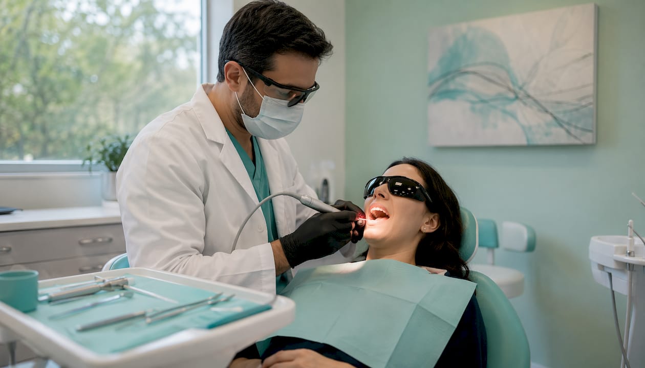
Flapless techniques are appropriate only in a narrow set of cases: adequate keratinized tissue, minimal osseous recontouring needed, and a thick/flat gingival phenotype. Laser-assisted gingival contouring (Er:YAG, diode) can be effective for soft-tissue-only cases but does not replace osseous surgery when bone removal is required. A CBCT-based APE classification helps identify which patients are candidates for flapless approaches.
Phenotype-driven technique selection:
- Thin/scalloped phenotype: favor minimal soft-tissue approaches or flapless techniques; full-thickness flap reflection risks fenestration and recession.
- Thick/flat phenotype: full-thickness flap with ostectomy/osteoplasty is predictable and well-tolerated.
Instrument checklist:
- Scalpel: #15 or #15C blade for precise esthetic incisions
- Periosteal elevator: for full-thickness flap reflection
- Piezoelectric unit or round carbide bur: for ostectomy and osteoplasty
- Periodontal curettes: for root surface debridement after osseous resection
- Laser (Er:YAG or diode): for soft-tissue-only flapless cases
- Suture: 5-0 or 6-0 chromic gut or polyglycolic acid for papilla preservation; interrupted or sling suture design
Practical tips for anesthesia, hemostasis, and suturing in the esthetic zone
Anterior esthetic cases demand more operative control than posterior crown lengthening. Small errors in hemostasis or suture placement show up directly in the smile.
Anesthesia:
- Use infiltration anesthesia (supraperiosteal) for maxillary anterior cases; avoid nerve blocks where possible to preserve lip proprioception and allow intraoperative smile assessment.
- Vasoconstrictor (1:100,000 epinephrine) is standard; wait a full 7–10 minutes after injection before incising to maximize hemostasis.
- For palatal tissue when needed, a greater palatine or nasopalatine block provides adequate coverage without distorting labial tissue.
Hemostasis:
- Epinephrine-containing local anesthetic is the primary hemostatic tool; supplemental infiltration into the papillae immediately before incision helps.
- Topical hemostatic agents (oxidized cellulose, collagen sponge) can be placed in the sulcus after flap reflection if oozing obscures the osseous landmarks.
- Avoid electrosurgery on marginal gingiva in thin phenotypes; the risk of recession and scarring outweighs the hemostatic benefit.
Suturing and papilla management:
- Interrupted sutures at each papilla tip maintain position and prevent the “black triangle” that opens when papillary tissue collapses.
- Sling sutures around individual teeth allow precise flap positioning without tension on the papillae.
- 5-0 or 6-0 resorbable sutures (chromic gut or polyglycolic acid) minimize tissue trauma; remove non-resorbable sutures at 7–10 days.
- Never place a suture through the papilla tip itself; pass through the base of the papilla to avoid ischemia and necrosis.
Common intraoperative pitfalls:
- Removing too little bone because the surgeon is referencing the current gingival margin instead of the planned restorative margin
- Raising the interdental papilla in esthetic cases, which leads to open embrasures
- Placing the flap too coronally after ostectomy, which allows tissue rebound and negates the bone removal
- Failing to reconfirm the margin-to-crest distance with a probe before suturing
When is it safe to place the final restoration after surgery?
Healing timeline and evidence-based timing
Wound healing after esthetic crown lengthening follows predictable phases, but the timeline for gingival margin stability is longer than most clinicians expect, especially in anterior esthetic cases.
Healing phases and tissue changes:
- Weeks 1–2: Initial clot organization, epithelial migration, and early granulation tissue formation; the gingival margin is edematous and positioned apically.
- Weeks 3–6: Connective tissue maturation; the margin begins to migrate coronally (rebound). This is the phase that catches clinicians off guard.
- Months 2–6: Continued collagen remodeling and sulcus re-establishment; the margin stabilizes, but thin phenotypes and extensive ostectomy cases may continue to shift.
Evidence-based timing recommendations:
- Functional/posterior cases: re-evaluate at a minimum of 6 weeks before placing final restorations. Prosthetic treatment should begin no earlier than six weeks following surgery.
- Esthetic anterior cases: a minimum of 3 months is suggested in many esthetic protocols, and gingival margin stability in high-smile esthetic cases often requires 4–6 months for full maturation.
- Thin phenotype, extensive ostectomy, or older patients: plan for the longer end of the range (5–6 months) and use provisional refinements to track margin position.
Statistic callout: The supracrestal tissue attachment averages approximately 2.04 mm (junctional epithelium ~0.97 mm + supracrestal connective tissue ~1.07 mm). Adding the mean sulcus depth (~0.69 mm) gives the practical 3 mm surgical landmark from desired margin to alveolar crest.
Follow-up schedule:
- 1 week: suture removal (non-resorbable), wound inspection
- 3–4 weeks: assess tissue rebound, adjust provisional if needed
- 6–8 weeks: re-evaluate margin position; photograph and compare to baseline
- 3–6 months: confirm margin stability before taking final impressions or scanning
Provisional restorations are not passive during this period. Adjust the provisional contours at each visit to guide the gingival margin toward the desired position. A provisional that is too bulky coronally will push the tissue apically; one that is too thin will allow coronal creep.
What complications should you prepare for after esthetic crown lengthening?
Prevention, recognition, and management
Most complications after esthetic crown lengthening are preventable with correct planning and technique. The ones that do occur are manageable when caught early.
Common complications and early signs:
- Recession beyond the planned level: gingival margin apical to the desired position at 6–8 weeks; often from thin phenotype, aggressive ostectomy, or flap placed too apically
- Tissue rebound (coronal creep): margin migrates coronally after surgery; most common when insufficient bone was removed; can negate the procedure
- Root sensitivity: exposed cementum after recession; managed with desensitizing agents, fluoride varnish, or dentin bonding
- Black triangles / open embrasures: loss of papillary height after flap reflection; more common when papillae were raised or when interproximal bone was aggressively reduced
- Esthetic mismatch: final crown proportions or gingival levels differ from the planned design; usually a planning failure, not a healing failure
Prevention strategies:
- Match technique to phenotype: thin/scalloped phenotypes need conservative soft-tissue approaches.
- Remove adequate bone: under-resection is the most common technical error and leads to rebound.
- Use the staged approach for complex cases to assess tissue response before committing to osseous surgery.
- Preserve the interdental papillae: do not raise them during flap reflection in esthetic cases.
Management algorithms:
- Early recession (6–8 weeks): monitor; adjust provisional to support tissue; consider connective tissue graft if recession exceeds 1 mm beyond the planned level and is not stabilizing.
- Continued rebound at 3 months: reassess bone removal; if the crest is within 2 mm of the margin, surgical revision with additional ostectomy may be required.
- Root sensitivity: apply 5% sodium fluoride varnish at each visit; consider dentin bonding agent if sensitivity persists beyond 3 months.
- Black triangles: hyaluronic acid injection into the papilla (off-label, emerging evidence) or restorative contouring of contact point position; surgical correction is rarely indicated.
- Esthetic mismatch: document with photographs, review the original plan, and determine whether the error was in planning (wrong margin position) or execution (inadequate bone removal or flap placement).
Red flags requiring re-evaluation or referral:
- Persistent mobility or bone loss beyond the surgical site
- Failure of the margin to stabilize after 6 months
- Patient dissatisfaction with esthetics that cannot be resolved with provisional adjustments
Document all complications, management steps, and patient communications in the chart. This is both a clinical and medicolegal requirement.
What are the alternatives when esthetic crown lengthening is not the right choice?
Not every gummy smile needs surgery, and not every short clinical crown needs bone removal. Knowing the alternatives makes you a better diagnostician and a more credible treatment planner.
Orthodontic intrusion:
Intrusion of over-erupted teeth reduces gingival display without removing tissue or bone. It is the preferred option when the etiology is dentoalveolar (teeth and supporting bone have erupted too far), particularly in younger patients where tissue remodeling is favorable. The limitation is time: orthodontic intrusion takes months, and the gingival tissue often follows the tooth unless surgical recontouring is performed afterward.
Prosthetic and restorative options:
Veneers or crowns can lengthen the apparent clinical crown by adding incisal length without touching the gingival margin. This works well when the crown is short due to wear and the gingival levels are already acceptable. The tradeoff is tooth preparation and the irreversibility of the restoration. For patients who refuse surgery, prosthetic lengthening is a reasonable compromise, but it does not address the underlying gingival display.
Lip repositioning surgery:
For patients whose excessive gingival display is driven by a hypermobile upper lip, lip repositioning reduces the muscle pull that elevates the lip during smiling. It is a soft-tissue procedure with a relatively short recovery and no osseous component. Results are generally stable, though some relapse is reported.
Botulinum toxin for hyperactive upper lip:
Botulinum toxin injected into the levator labii superioris reduces lip elevation and decreases gingival display. It is reversible, minimally invasive, and useful for patients who want to preview the esthetic result before committing to surgery. The effect lasts 3–6 months, so it is a maintenance treatment rather than a permanent solution.
Combining modalities:
The most demanding esthetic cases often require more than one approach. Orthodontic intrusion followed by minimal esthetic crown lengthening reduces the amount of bone removal needed. Prosthetic lengthening combined with conservative gingival contouring avoids aggressive osseous surgery in thin-phenotype patients. The key is sequencing: complete orthodontic treatment before surgery, and complete surgery before final restorations.
A clinical checklist for predictable esthetic crown lengthening outcomes
Preoperative, intraoperative, and postoperative coordination
This checklist is designed for use by both the periodontist and the restorative clinician. Run through it at the treatment planning appointment and again on the day of surgery.
Preoperative checklist:
- Full-face and retracted photographs (rest and full smile) obtained and reviewed
- Diagnostic wax-up or digital smile design completed and approved by patient
- Provisional restoration fabricated; surgical guide derived from provisional
- Periodontal charting, bone sounding, and CEJ-to-crest measurements documented
- CBCT reviewed if root morphology, bone thickness, or APE classification is uncertain
- Keratinized tissue width confirmed (≥2 mm post-surgery target)
- Informed consent completed: recession risk, sensitivity, black triangle risk, timing of final restoration, possibility of staged revision
Intraoperative targets:
- Seat surgical guide before first incision; confirm incision line matches planned gingival zenith
- Reflect buccal flap only; do not raise interdental papillae
- After ostectomy, confirm ~3 mm from planned restorative margin to alveolar crest with a periodontal probe
- Adapt flap to CEJ level or slightly coronal; no tension on papillae
- Place provisional restoration immediately; confirm it does not impinge on the healing supracrestal tissue
Postoperative timeline and contingency triggers:
- 1 week: suture removal; wound inspection; photograph
- 3–4 weeks: assess rebound; adjust provisional contours
- 6–8 weeks: re-evaluate margin position; if rebound >1 mm, reassess bone removal adequacy
- 3 months: photograph and compare to baseline; if margin is stable, begin restorative planning
- Delay trigger: if the margin has not stabilized by 4 months, extend the provisional phase and re-evaluate at 5–6 months before taking final impressions
Clinician communication template (periodontist to restorative clinician):
Pro Tip: Photograph the provisional in place at every follow-up visit and overlay the images digitally. Margin migration of even 0.5 mm is visible in a side-by-side comparison and gives you objective data to decide when the tissue has stabilized.
Key Takeaways
Esthetic crown lengthening requires restoration-driven planning, phenotype-matched technique selection, and a minimum of 3–6 months of tissue maturation before placing definitive restorations in the esthetic zone.
| Point |
Details |
| Plan to the final margin |
Bone removal is dictated by the desired restorative margin, not the current gingival level. |
| Use the 3 mm surgical landmark |
Maintain approximately 3 mm from the planned margin to the alveolar crest to preserve supracrestal tissue attachment (average 2.04 mm + mean sulcus depth 0.69 mm). |
| Stage complex cases |
Staged surgery with provisional management reduces open embrasures and margin unpredictability. |
| Match technique to phenotype |
Thin/scalloped phenotypes need conservative approaches; thick/flat phenotypes tolerate full-thickness flaps with ostectomy. |
| Allow adequate maturation |
Wait 3–6 months for esthetic anterior cases before final restorations; 6 weeks is the minimum for posterior functional cases only. |
| Build skills with Onewd |
Onewd’s hands-on training courses offer tiered surgical and restorative CE pathways for clinicians developing ECL competency. |
The mistake most clinicians make is not the surgery itself
There is a pattern that shows up repeatedly when clinicians are learning esthetic crown lengthening: they perform technically adequate surgery and then rush the restorative phase. The tissue looks stable at six weeks, the patient is asking for their final crowns, and the path of least resistance is to proceed. Three months later, the gingival margin has crept coronally, the crown margins are exposed, and the esthetic result is compromised.
The harder lesson to teach is that the provisional phase is not waiting time. It is active treatment. The provisional restoration is shaping the tissue, guiding the margin, and giving you real-time information about where the final margin will actually sit. Clinicians who treat it as a placeholder miss the most valuable diagnostic window in the entire case.
The second pattern is under-resection of bone. It feels conservative to remove less, but insufficient ostectomy is the primary driver of tissue rebound. The tissue will always try to re-establish its supracrestal attachment dimensions. If you have not given it enough room apically, it will take that room coronally, at the expense of your surgical result. The 3 mm landmark from planned margin to crest is not a suggestion; it is the minimum required for the tissue to stabilize where you want it.
The third pattern is poor interdisciplinary communication. The periodontist and the restorative clinician need to agree on the final margin position before the first incision, not after. Cases where this conversation happens post-surgically almost always result in a mismatch between what was surgically achieved and what the restoration requires.
The fix for all three is the same: slow down the planning phase, speed up nothing else.
Advance your esthetic crown lengthening skills with Onewd
Esthetic crown lengthening sits at the intersection of periodontics and restorative dentistry, and the gap between knowing the technique and executing it predictably in live patients is real. Reading the literature gets you the framework; hands-on training with mentorship gets you the confidence to apply it.
Onewd offers tiered surgical and cosmetic dentistry courses that build exactly the competencies this procedure demands: flap design, osseous recontouring, provisional management, and interdisciplinary case coordination. Courses range from on-demand modules for foundational technique review to live patient hands-on programs where you perform procedures under expert guidance. CE credits are available across formats, so you can satisfy licensure requirements while building real clinical skill. The mentorship-driven model means you get feedback on your actual case decisions, not just lecture content.
If you are ready to move from understanding the principles to executing them predictably, browse Onewd’s upcoming hands-on courses and find the format that fits your schedule and learning goals.
Useful sources for further reading
The following peer-reviewed references and clinical resources support the evidence base presented in this guide. Clinicians are encouraged to consult primary literature when documenting consent and planning complex cases.
- Staged esthetic crown lengthening: Classification and guidelines for periodontal-restorative therapy — Primary source for restoration-driven planning, staging rationale, supracrestal tissue attachment measurements, and the 3 mm surgical landmark.
- Clinical considerations for crown lengthening (PMC/NIH) — Evidence summary supporting delayed restorative timing in esthetic cases and supracrestal tissue attachment maintenance.
- Guiding esthetic crown lengthening: A CBCT-based modified classification of altered passive eruption — CBCT-based APE classification; supports flapless vs. open-flap technique selection.
- Experts: etiology and diagnosis of excessive gingival display and APE considerations — Covers differential diagnosis of gummy smile, supracrestal tissue attachment terminology shift, and phenotype-driven planning.
- Crown Lengthening Revisited (University of Michigan) — Comprehensive decision-tree resource for esthetic vs. functional crown lengthening technique selection.
- Biologic width and its importance in periodontal and restorative dentistry (PMC) — Reviews biologic width concepts, margin placement rules, and healing timelines after crown lengthening.
- Biologic Width Following Different Crown Lengthening Procedures: A Six-Month Follow-Up Study (PMC) — Clinical comparison of gingivectomy vs. apically positioned flap with ostectomy for biologic width re-establishment at 6 months.
- Aesthetic crown lengthening: Classification, biologic rationale, and treatment planning (Lee, Pract Proced Aesthet Dent) — Classic classification system (Type I–IV) and surgical guide rationale; foundational reference for staging and technique selection.
- Concepts and considerations for surgical crown lengthening (Decisions in Dentistry) — Practical presurgical considerations including keratinized tissue width and crestal bone position for flap design.
Recommended