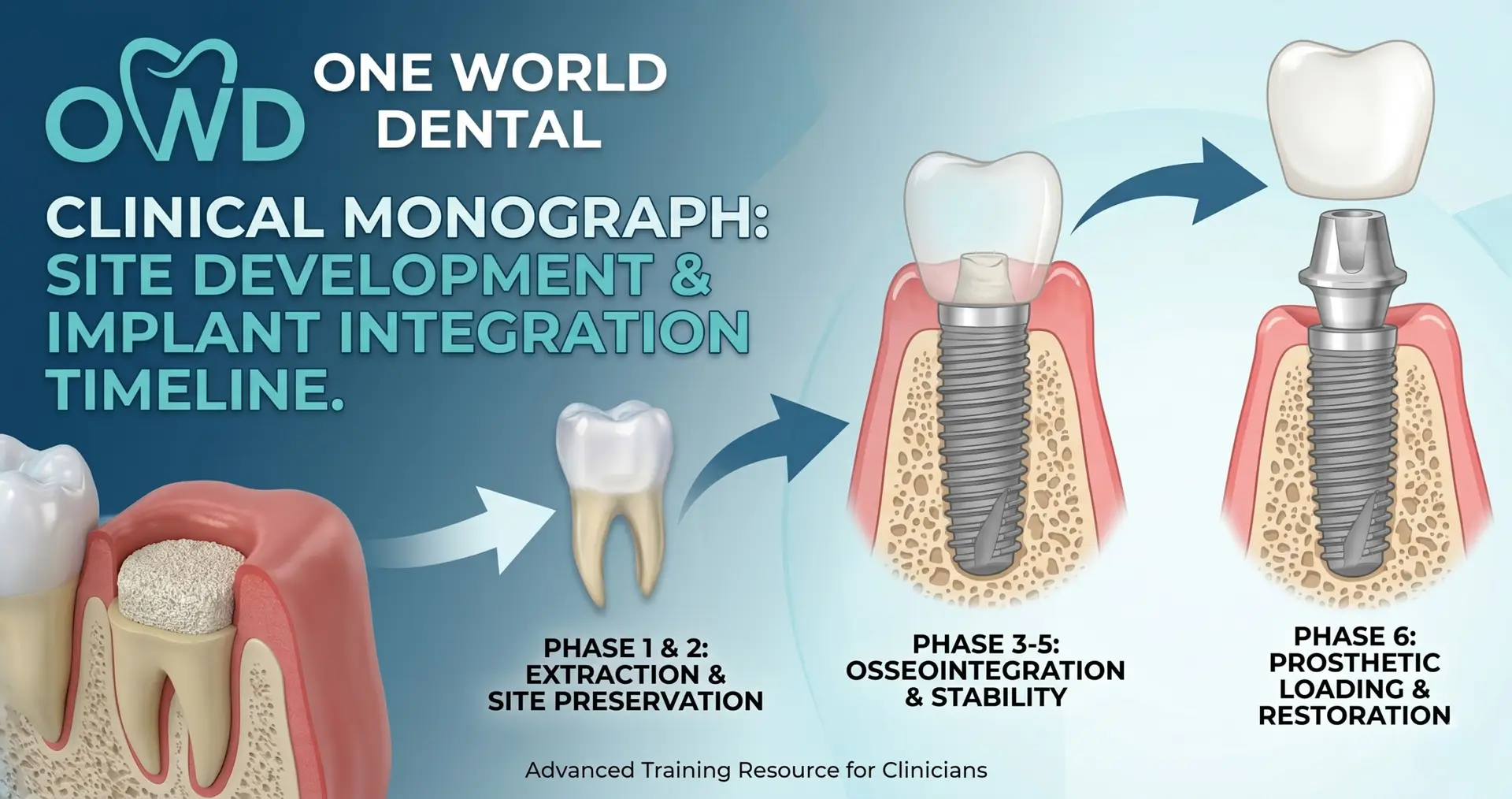
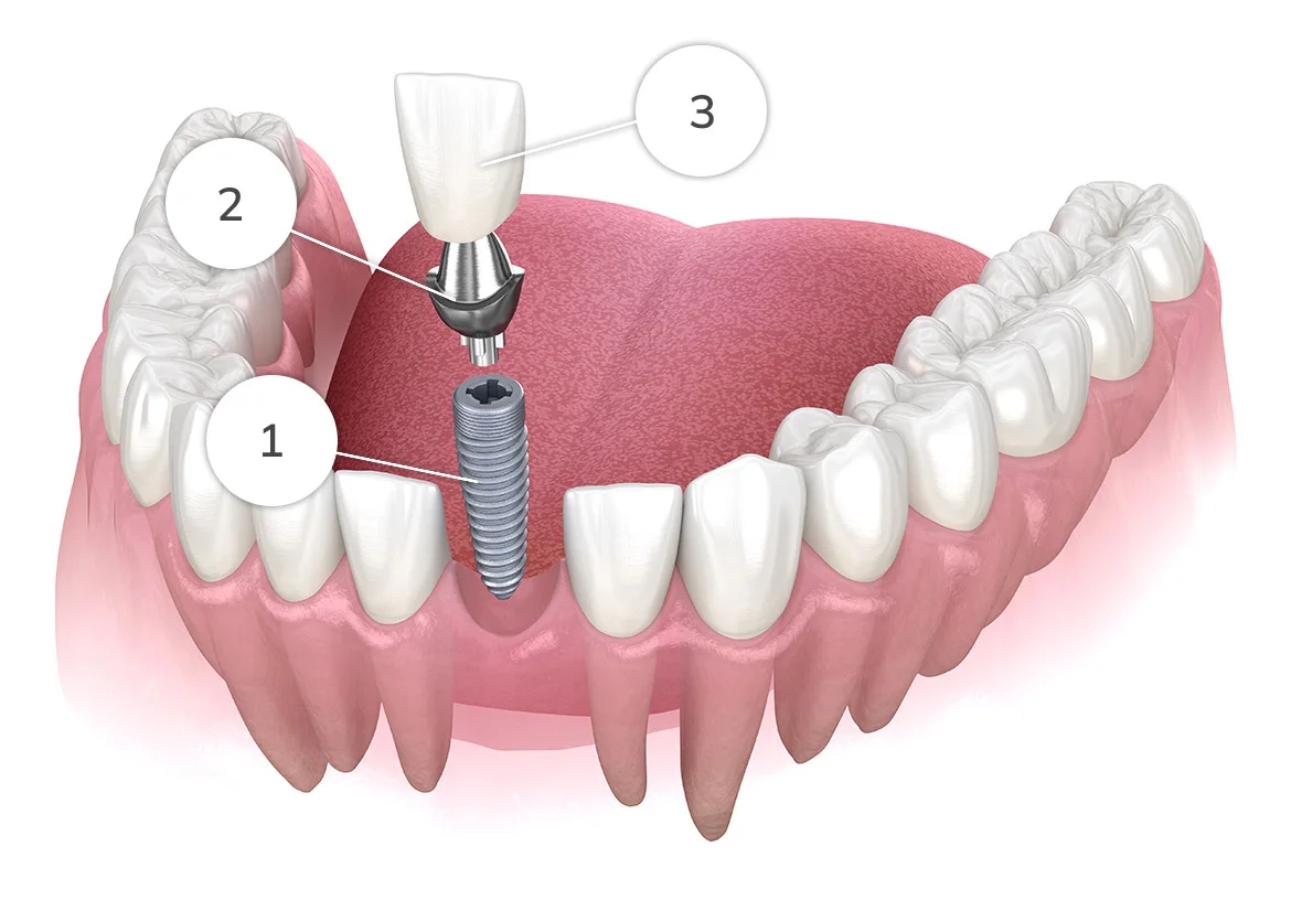
The predictability of the long-term endosseous integration of titanium and zirconia fixtures is predicated on the fastidious execution of Site Development. This protocol transitions the clinician from reactive tooth replacement to proactive alveolar reconstruction. At One World Dental, our surgical philosophy prioritizes the maintenance of the cortical plates and the optimization of the regenerative “Golden Window” to mitigate the 40-60% post-extraction ridge resorption typically observed within the first 12 months.
Pre-Surgical Diagnostics: The Digital Blueprint
CBCT Volumetric Analysis (D0367)
Practitioners must utilize high-resolution CBCT to assess the Lekholm and Zarb bone classification. Site development strategies differ significantly between Type II (dense cortical/thick trabecular) and Type IV (thin cortical/low-density trabecular) bone.
Clinical Gap: Mapping the Vascular Supply. Identifying the location of the canalis sinuosus or the mandibular canal with sub-millimeter precision is non-negotiable for 3D guided surgery.
Systemic Biomarkers and Host Modulation
Successful site development is a biological endeavor, not just a mechanical one.
Vitamin D3 (25-hydroxy): Patients with serum levels below $30ng/mL$ exhibit increased rates of early implant failure due to impaired osteoblast activity.
HbA1c Levels: For diabetic patients, maintaining a level $<7.0\%$ is critical for microvascular health at the surgical site.
CD4+ T-cell Counts: In immunocompromised patients, the timeline for maturation must be extended by 25-40%.
Atraumatic Extraction and Alveolar Ridge Preservation
Physics-Aided Exodontia
To preserve the buccal plate—crucial for esthetic “pink porcelain” results—the clinician should employ periotomes or piezosurgery instead of traditional forceps. The goal is the complete severance of the periodontal ligament (PDL) without luxation-induced fractures of the thin labial cortical bone.
Socket Preservation Protocols (D7953)
Particulate Allograft vs. Xenograft: While allografts offer faster remodeling, xenografts (bovine-derived) provide superior long-term volume stability due to their slower resorption rate.
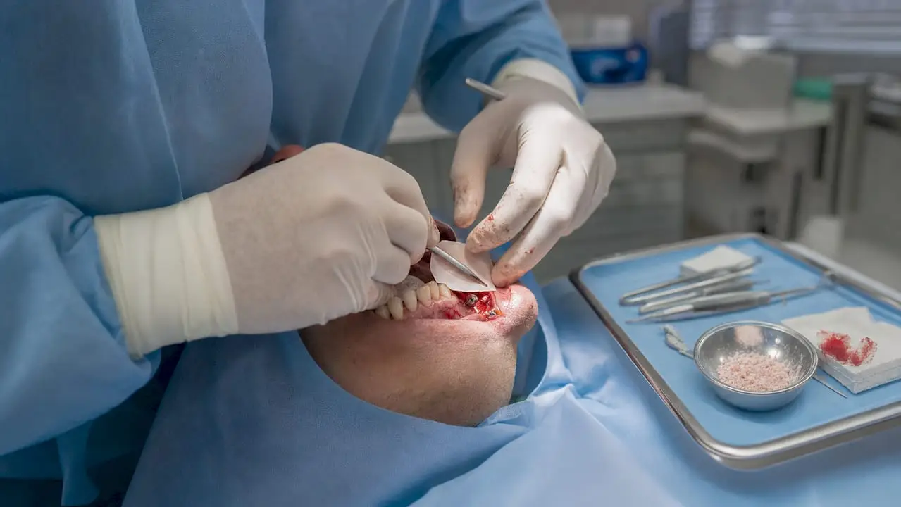
The “Iceberg” Technique: Clinicians should over-fill the socket slightly and secure the site with a non-resorbable dPTFE membrane. This “open-membrane” technique facilitates secondary intention healing while excluding epithelial downgrowth, eliminating the need for primary closure which often displaces the mucogingival junction.
Advanced Site Augmentation: Beyond the Socket
Vertical and Horizontal Ridge Augmentation
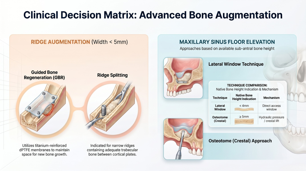
When the residual ridge width is $<5mm$, specialized augmentation is required:
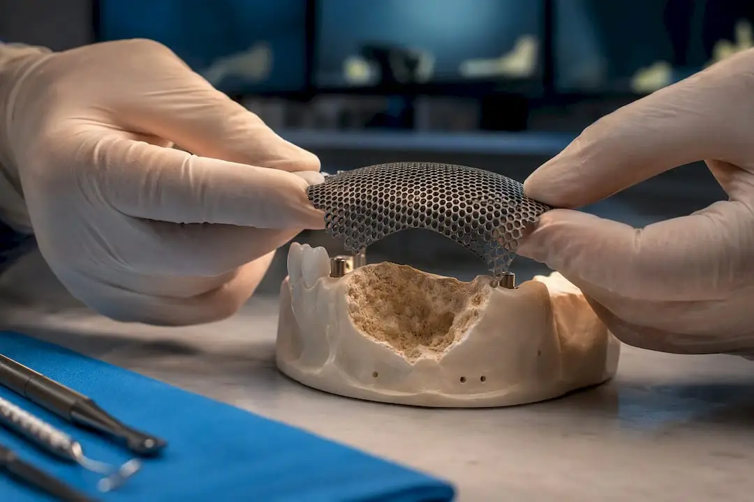
Guided Bone Regeneration (GBR): Utilizing titanium-reinforced membranes (dPTFE) to maintain space.
Ridge Splitting: Indicated for narrow ridges with adequate trabecular bone between cortical plates.
Maxillary Sinus Floor Elevation (D7911/D7912)
In the posterior maxilla, sub-antral bone height is often insufficient.
Lateral Window Technique: Indicated for $<4mm$ of native bone.
Osteotome (Crestal) Approach: Indicated when $\geq5mm$ of bone exists, utilizing hydraulic pressure to lift the Schneiderian membrane.
Implant Placement: Surgical Precision
Primary Stability and Insertion Torque (IT)
The clinical goal is an IT of 35-45 Ncm.
Undersized Osteotomy: In Type IV bone, preparing a site smaller than the fixture diameter leverages “bone condensing” to increase primary stability.
Resonance Frequency Analysis (RFA): Use ISQ (Implant Stability Quotient) values to establish a baseline for secondary stability.
The Osseointegration Gap: Secondary Stability
The Stability Dip
Between weeks 2 and 4, primary stability (mechanical) decreases while secondary stability (biological) is not yet established.
Clinical Nuance : This is the high-risk “vulnerability window.” Practitioners must ensure the provisional restoration is in non-functional immediate transition (no occlusal contact in centric or eccentric movements) to avoid micro-motion exceeding $150\mu m$, which leads to fibrous encapsulation rather than osseointegration.
Surgery and Soft Tissue Management
The Roll Flap and Apically Positioned Flap
At the time of abutment connection (D6056), the clinician must ensure a minimum of 2mm of keratinized mucosa (KM) circumferentially. If KM is lacking, a free gingival graft (FGG) or connective tissue graft (CTG) should be performed to prevent future peri-implantitis.
Prosthetic Loading and Long-Term Maintenance
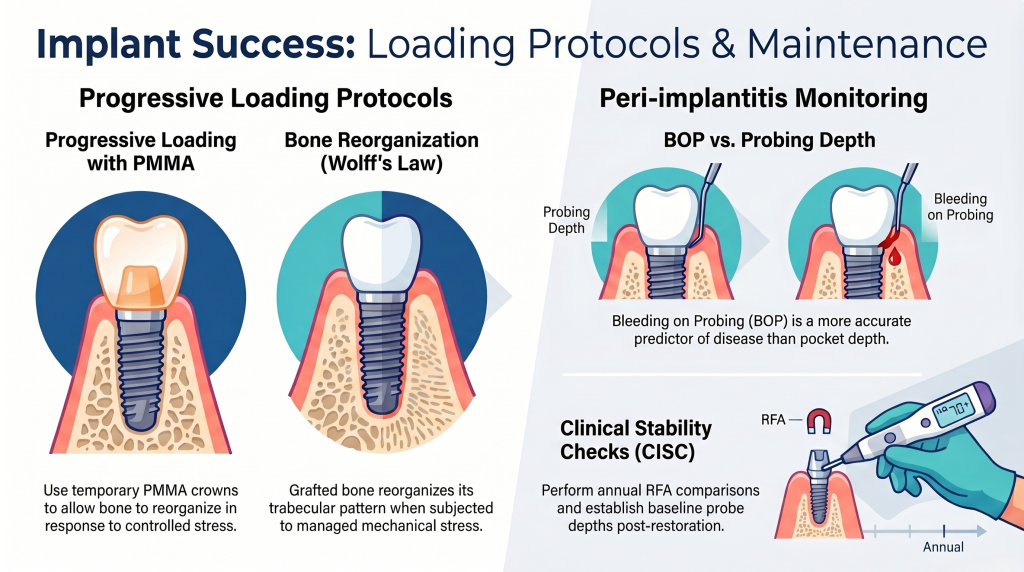
Progressive Loading Protocols
In cases of grafted bone, “progressive loading” using a temporary PMMA crown allows the bone to reorganize its trabecular pattern in response to controlled mechanical stress (Wolff’s Law).
Probing Depth: Establishing a “baseline” probe depth post-restoration. Bleeding on Probing (BOP) is a more accurate predictor of disease than pocket depth in implants.
People Also Ask (Clinical FAQ)
What is the optimal healing time for an autogenous block graft versus particulate graft? Autogenous blocks typically require 4–5 months, whereas particulate grafts in a protected socket environment can often be implanted at 3.5–4 months depending on the graft-to-native bone ratio.
When is “Immediate Placement” (D6010) contraindicated? Immediate placement is contraindicated in the presence of active purulent exudate, lack of at least 3mm of bone apical to the extraction socket for primary stability, or a compromised buccal plate.
How does PRF (Platelet-Rich Fibrin) influence the site development timeline? L-PRF accelerates soft tissue healing and initial angiogenesis. While it doesn’t necessarily shorten the 4-month bone maturation period, it significantly reduces the “stability dip” by promoting faster osteoblast migration.
What is the CDT coding for a Sinus Lift with an implant? Use D7911 for the sinus lift (lateral) or D7912 (crestal) in conjunction with D6010 for the surgical placement of the implant body.
How do bisphosphonates affect the site development timeline? Patients on oral bisphosphonates for $>3$ years require a “drug holiday” (consulting with their MD) and a C-terminal telopeptide (CTX) test to ensure bone turnover is sufficient for healing ($>150 pg/mL$).
Conclusion: Maximizing Clinical Outcomes
Site development is a disciplined approach to biological engineering. By adhering to these advanced surgical protocols and respecting the physiological timelines of bone remodeling, clinicians can provide restorations that rival the longevity of natural dentition.
For complex multidisciplinary cases involving ridge augmentation or advanced sinus surgery, One World Dental serves as a premier referral hub for practicing clinicians. We utilize the latest in PRF growth factors and CBCT-guided surgery to ensure every site is optimized for maximum stability.
Educational disclaimer: Content is provided for professional education and is not a substitute for a clinician's independent judgment, applicable standards, manufacturer instructions, or relevant laws and regulations.
For clinicians: a defect-based ridge augmentation guide that matches technique to anatomy, explains mesh and CAD/CAM workflows, plans 4–6 month staging,...