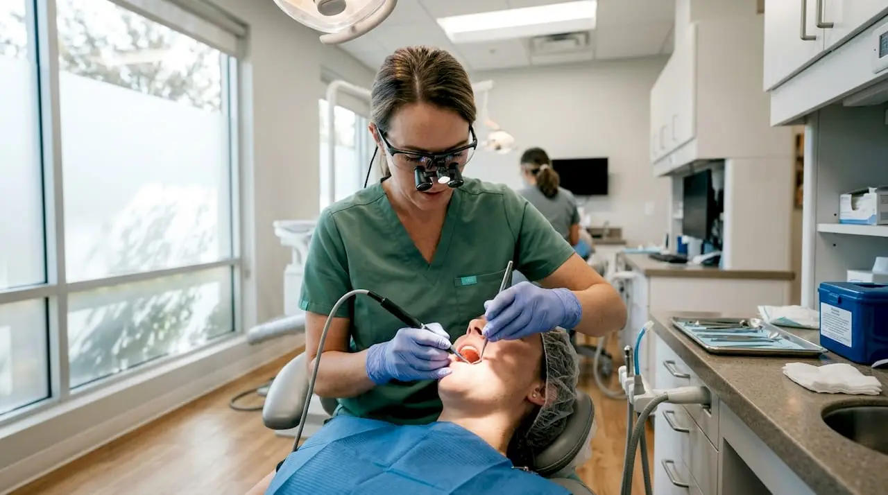
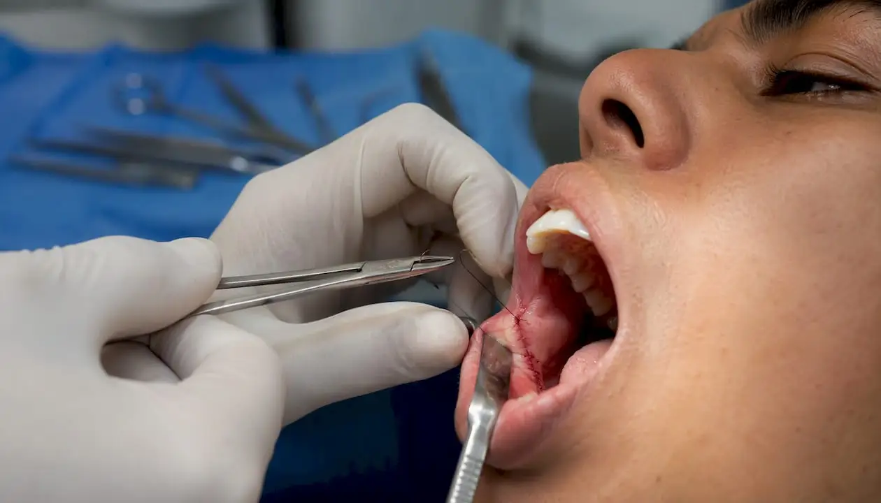
As a modern clinician, the paradigm of tooth removal has fundamentally shifted. The days of aggressive, high-force extractions are gone. Today’s standard of care focuses entirely on atraumatic extraction, ridge preservation, and hard/soft tissue management to set the stage for successful future implantology.
At One World Dental, our surgical training modules emphasize that a dental extraction is no longer just the removal of a non-rehabitable tooth—it is the first phase of reconstructive implant surgery.
Below, we break down the clinical foundations, advanced instrumentation, and biological protocols we teach to help clinicians transition from traditional exodontia to modern, predictable surgical extractions.
Shifting the Mindset: Atraumatic Exodontia vs. Traditional Removal
Traditional exodontia relied heavily on the mechanical advantages of forceps and elevators to expand the alveolar housing. While effective for simple cases, this approach frequently results in the fracture of delicate buccal plates, leading to significant ridge resorption.
Modern surgical extraction aims to preserve the periodontium entirely. By understanding how to strategically weaken the root-to-bone interface without stressing the surrounding housing, you can routinely leave the socket walls perfectly intact.
Advanced Clinical Mapping and CBCT Diagnostics
Predictable surgery starts before making an incision. We teach clinicians how to fully leverage 3D Cone Beam Computed Tomography (CBCT) to map out high-risk anatomical structures:
Nerve Mapping: Isolating the exact path of the inferior alveolar nerve (IAN) relative to impacted roots or complex mandibular root morphologies.
Sinus Proximity: Evaluating the floor of the maxillary sinus to prevent unintended oroantral communications (OAC) during upper molar extractions.
Root Morphology: Identifying dilacerations, hypercementosis, or multi-rooted teeth that dictate a planned tooth-sectioning approach rather than a force-based delivery.
Beyond the Forceps: Advanced Instrumentation
To achieve truly atraumatic results, a clinician’s armamentarium must evolve beyond standard elevators. In our hands-on One World Dental courses, we train doctors on the precise utilization of:
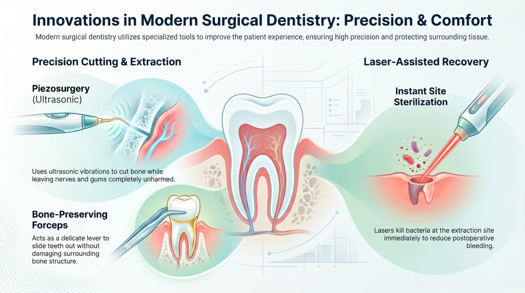
Periotomes and Luxators: Used to carefully sever the periodontal ligament (PDL) along the long axis of the tooth, minimizing lateral pressure on the cortical bone.
Physics Forceps: Utilizing a unique biomechanical design (a bumper acting as a fulcrum on the alveolus and a single beak engaging the lingual/palatal root surface), these instruments apply a constant, steady rotational force that utilizes the body’s viscoelastic property to allow the tooth to naturally “pop” out.
Piezosurgery (Ultrasonic Surgery): When bone removal or troughing is required, piezoelectric inserts allow for selective cutting of hard tissues while completely sparing adjacent soft tissues, nerves, and sinus membranes.
Biologics and Hard Tissue Grafting: Socket Preservation
A modern extraction is incomplete without a plan for managing the empty alveolus. Without intervention, the buccal plate can lose up to 40-60% of its volume within the first year.
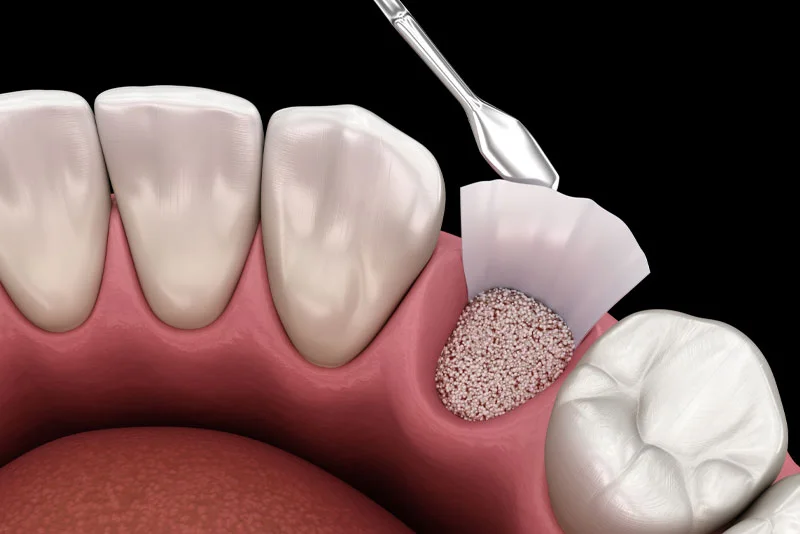
Socket Preservation Protocols: Packing the thoroughly debrided socket with an appropriate bone particulate (allograft, xenograft, or synthetic scaffold) to provide a matrix that supports new bone formation and prevents ridge collapse.
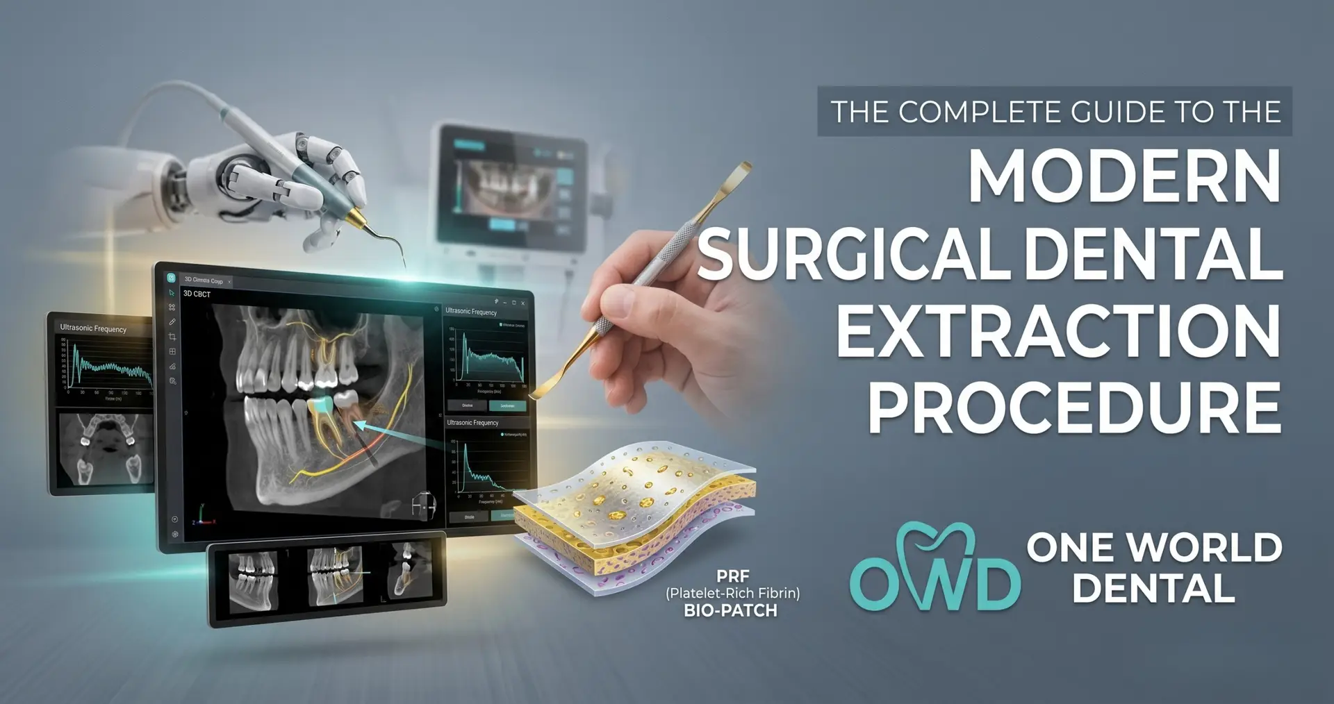
Platelet-Rich Fibrin (PRF): In our advanced clinical modules, we guide doctors through the process of venipuncture and centrifugation to create autologous PRF. Placing a PRF plug over the graft site introduces highly concentrated growth factors and cytokines that dramatically accelerate soft tissue closure, improve angiogenesis, and drastically reduce the incidence of alveolar osteitis (dry socket).
Post-Operative Management, Complications, and Clinical Documentation
The modern surgical extraction workflow doesn’t end when the final suture is tied. As clinical educators, we emphasize that comprehensive post-operative management and airtight documentation are vital for both patient outcomes and risk mitigation.
Evidence-Based Post-Op Protocols: Transitioning away from over-prescribing opioids toward highly effective, scheduled multimodal analgesia (such as alternating ibuprofen and acetaminophen). This manages acute inflammatory pain effectively while prioritizing patient safety.
Managing Complications Safely: Equipping your clinical team with clear protocols for handling secondary bleeding, delayed healing, or suspected infections. Providing patients with clear, written, and verbal instructions reduces emergency after-hours calls and ensures early intervention if a complication arises.
Defensive Clinical Charting: Your surgical notes must be detailed and objective. Ensure your documentation explicitly notes the presence/absence of the buccal plate post-extraction, the specific biomaterials used for socket preservation (including lot numbers and expiration dates), the type and count of sutures placed, and the patient’s understanding and acceptance of post-operative risks.
Frequently Asked Questions (Clinical FAQ)
When should I transition from a closed (simple) extraction to an open (surgical) extraction?
The transition should happen early in the procedure, not as a last resort after hours of struggling. If you experience a lack of progress after 5–10 minutes of controlled luxation, if the crown fractures at or below the bone crest, or if pre-operative CBCT imaging reveals highly divergent or dilacerated roots, you should immediately pivot to an open approach. Raising a clean flap and selectively guttering bone or sectioning the tooth saves time, preserves the buccal plate, and significantly reduces post-operative patient discomfort compared to prolonged, forceful instrumentation.
What is the clinical criteria for deciding between an allograft, xenograft, or synthetic material for socket preservation?
The choice depends heavily on your treatment timeline and restorative goals:
Allograft (human tissue): Offers rapid turnover and remodeling, typically making the site ready for implant placement within 3–4 months. It is ideal for standard implant timelines. Xenograft (bovine/porcine tissue): Remodels much slower, maintaining structural volume over a longer period. This is highly effective when implant placement is delayed (e.g., 6+ months) or when maximum space maintenance is needed in the aesthetic zone. Synthetic materials: Provide a highly predictable, osteoconductive matrix free of biological tissue risks, though remodeling rates vary widely by formulation.
Can I place Platelet-Rich Fibrin (PRF) directly over a bone graft without a traditional barrier membrane?
Yes, in many intact socket scenarios, an advanced PRF (A-PRF) matrix can act as both the biological stabilizer and the protective barrier. Because PRF contains dense fibrin networks packed with growth factors and leukocytes, it promotes incredibly rapid soft tissue epithelialization (often within 7 days). However, if you are treating a severe buccal wall defect or performing guided bone regeneration (GBR) where long-term space maintenance is required to prevent soft tissue downgrowth, a long-lasting resorbable or non-resorbable membrane is still clinically necessary. exercise for at least 3-5 days.
How much buccal bone loss is acceptable before immediate implant placement is contraindicated?
Generally, immediate implant placement requires a minimum of 2 mm of intact buccal bone thickness to prevent long-term soft tissue recession and aesthetic failure. If the buccal plate is completely missing or compromised greater than 3–4 mm apically from the crest, an immediate implant loses its predictable bone support. In these compromised scenarios, we train clinicians to favor a staged approach: perform a thorough atraumatic extraction, execute predictable socket preservation, and allow 4–6 months of solid hard-tissue regeneration before placing the fixture.
What is the most effective mechanical strategy to avoid fracturing the buccal plate during upper molar extractions?
Strategic tooth sectioning is your absolute best defense. Trying to deliver an intact upper molar often forces the divergent buccal roots to act as levers against the thin maxillary cortical bone. By utilizing a high-speed surgical handpiece to segment the tooth into three distinct, single-rooted sections (separating the mesiobuccal, distobuccal, and palatal roots), you can luxate each individual root along its own path of least resistance, leaving the structural anatomy of the socket completely uncompromised.
Elevate Your Clinical Skillset with One World Dental
Mastering these advanced surgical protocols requires more than just reading the literature—it requires hands-on, expert-guided mentorship.
Whether you are looking to confidently tackle impacted wisdom teeth, integrate PRF biologics into your daily practice, or master ridge preservation before placing implants, One World Dental’s tiered learning pathways provide the rigorous clinical training you need to excel.
Educational disclaimer: Content is provided for professional education and is not a substitute for a clinician's independent judgment, applicable standards, manufacturer instructions, or relevant laws and regulations.