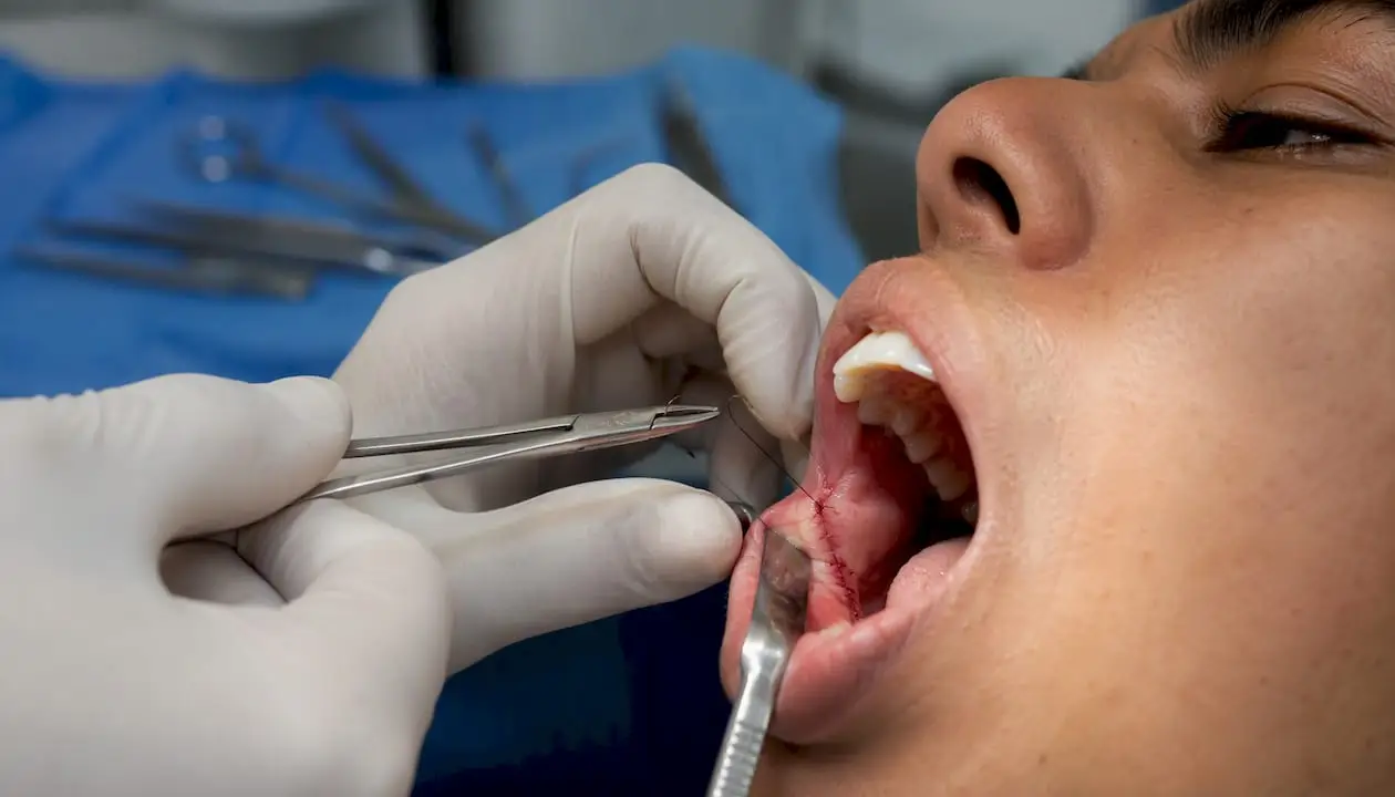
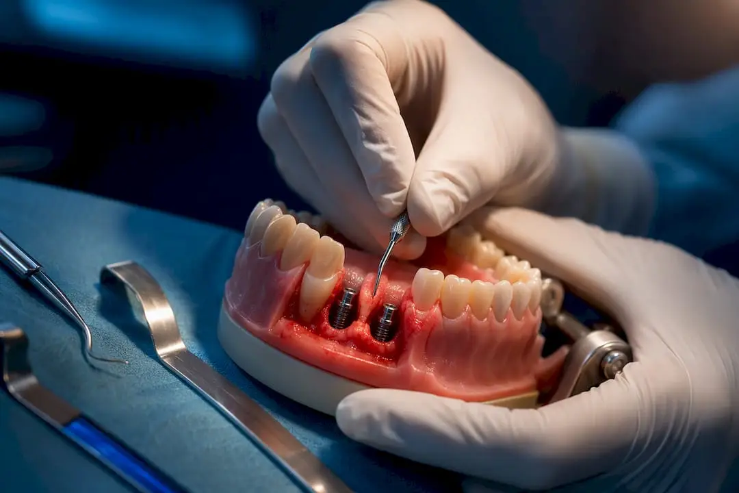
Dental electrosurgery is defined as the use of high-frequency radiofrequency energy to simultaneously incise and coagulate oral soft tissue during surgical procedures. Also called electrocautery or radiosurgery in clinical settings, the technique gives you precise tissue control that a scalpel alone cannot match. The closed-circuit design delivers current through an active electrode tip, through the patient, and back through a neutral electrode, completing the circuit without systemic risk. Core applications include gingivectomy, gingival recontouring, frenectomy, abscess drainage, and impacted tooth exposure, covering periodontics, restorative dentistry, orthodontics, and oral surgery. Understanding the technology, modes, and safety protocols is what separates a practitioner who uses this tool confidently from one who avoids it.
What are the main clinical applications of dental electrosurgery?
Electrosurgery covers a wider range of soft tissue procedures than most clinicians initially realize. The technique applies across four major dental specialties, each with distinct procedural goals.
Common clinical indications include:
- Gingivectomy and gingival recontouring: Reshaping hyperplastic or excess gingival tissue for periodontal health or esthetic correction. Hemostasis during the cut keeps the field dry and visible throughout.
- Frenectomy: Releasing labial or lingual frenum attachments. Electrosurgery reduces bleeding significantly compared to scalpel excision, which matters in a tight anatomical space.
- Incision and drainage of abscesses: The electrode both opens the lesion and sterilizes the wound margin simultaneously. High-frequency energy denatures tissue proteins, which reduces postoperative inflammation risk.
- Crown lengthening and tissue retraction: Removing gingival tissue to expose more tooth structure before restorative work. A dry, retracted margin directly improves impression accuracy.
- Exposure of impacted teeth: Orthodontic and surgical cases benefit from the clean, bloodless access electrosurgery provides.
- Operculectomy and epulis removal: Removing soft tissue flaps or benign lesions with controlled depth and minimal collateral spread.
Surgical gingivectomy remains the most common approach, performed in 61.25% of cases compared to electrocautery at 31.55% and laser gingivectomy at 7.50%. That distribution shows electrosurgery holds a substantial and growing share of clinical practice.
Pro Tip: For crown lengthening before final impressions, use electrosurgery to retract and contour in a single pass. You eliminate the need for retraction cord in many cases, saving chair time and reducing patient discomfort.
How do dental electrosurgery units work?
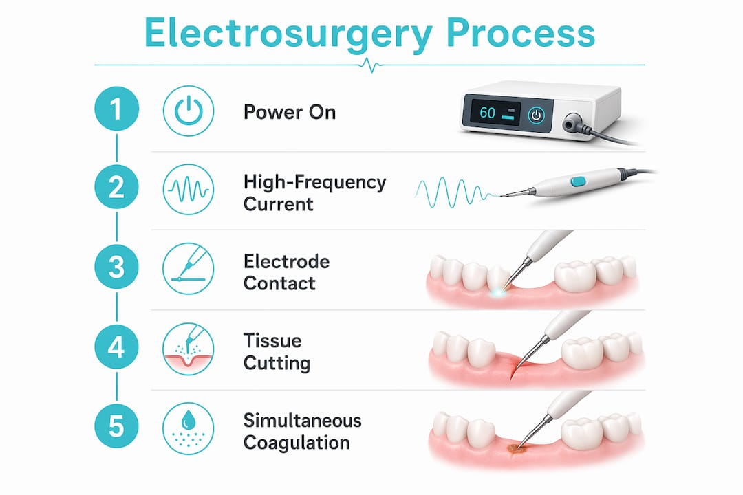
The core of any electrosurgical unit is a high-frequency generator that produces alternating current in the radiofrequency range, typically between 1 MHz and 4 MHz. At these frequencies, the current passes through tissue without triggering neuromuscular stimulation. The circuit requires both an active electrode at the surgical site and a neutral electrode, usually a conductive bracelet worn by the patient, to complete the loop safely.
Operating modes and their clinical effects
Electrosurgical units offer four primary modes, each producing a distinct tissue response:
- Cut (fully filtered): Continuous waveform output. Produces clean incisions with minimal lateral heat spread. Use this for gingivectomy and frenectomy where tissue architecture matters.
- Coagulation: Pulsed waveform output. Denatures surface proteins to arrest bleeding. Higher lateral heat than cut mode, so limit contact time.
- Blended: Combines cut and coagulation waveforms. Useful when you need simultaneous incision and hemostasis, such as during crown lengthening in vascular tissue.
- Fulguration: Non-contact coagulation using sparking. Applied for surface lesion destruction or superficial hemostasis without direct tissue contact.
Electrode tips and selection
Electrode tip geometry determines the precision and depth of the cut. Needle and wire loop tips produce fine incisions for recontouring work. Ball tips, such as the FC10N, are designed for coagulation. Blade tips work for broader excisions. Specific tips like the I22S and I22CA are optimized for gingivectomy, while ball electrodes serve coagulation tasks. Color coding on tip families helps you select the right geometry quickly at the unit.
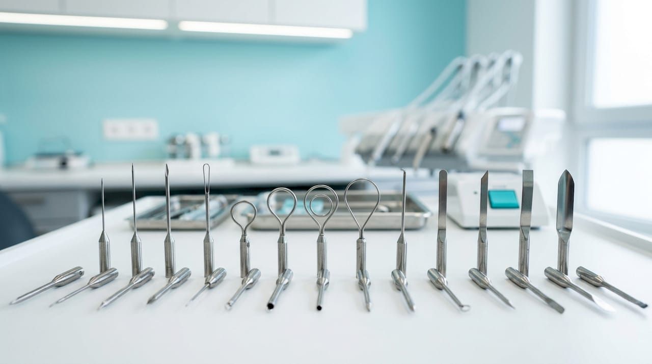
Power output also matters. Units like the Servotome deliver up to 30 W, with settings adjusted per tissue density and procedure type. Lower settings reduce lateral heat; higher settings cut faster but increase thermal spread risk.
Pro Tip: Start at the lowest effective power setting and increase only if tissue drag occurs. Cutting with excess power is the most common cause of unintended thermal damage in electrosurgery.
Neutral electrode safety design
Conductive bracelet neutral electrodes have replaced older grounding plates in modern units. The bracelet design improves patient hygiene, reduces setup time, and eliminates the risk of pad burns from poor skin contact. Always inspect the bracelet and cable before each procedure.
What are the advantages and limitations of electrosurgery vs. other methods?
Electrosurgery’s unique ability to simultaneously cut and coagulate enhances operative visualization. That is its primary clinical advantage over the conventional scalpel. A dry, clear field reduces procedural errors and shortens operating time.
Advantages
- Superior hemostasis: Electrosurgery controls bleeding in real time, which scalpel techniques cannot do without additional hemostatic agents.
- Wound sterilization: High-frequency energy sterilizes the wound margin as it cuts, reducing postoperative inflammation in sites like post-extraction wounds.
- Operative field clarity: Continuous hemostasis keeps the surgical site visible throughout the procedure.
- Affordability: Electrosurgical units cost significantly less than diode or Er:YAG laser systems, making the technology accessible for general practice.
- Versatility: One unit covers multiple procedures across specialties without changing platforms.
Limitations
Electrosurgery entails longer healing times and more postoperative discomfort than diode laser treatments. Diode lasers provide faster tissue healing and less postoperative pain, which matters for patient experience and same-day follow-up calls. Anesthesia requirements also differ: electrosurgical gingivectomy typically requires 3–5 anesthetic vials, while laser-assisted procedures require only 1–3. That gap affects both patient comfort and procedure cost.
Technique selection should balance lesion extent, tissue biotype, and patient affordability rather than default to the most advanced available technology. Thin, fragile tissue biotypes respond poorly to excess lateral heat, making laser or scalpel preferable in those cases. For thick, vascular tissue where hemostasis is the primary concern, electrosurgery remains the most practical choice.
What are best practices and safety protocols for dental electrosurgery?
Safe and effective electrosurgery depends on technique discipline as much as equipment quality. The following protocols reduce complication risk and improve outcomes.
- Use short-trigger activation with continuous tip movement. Short bursts with constant motion prevent heat accumulation at any single point. Holding the electrode stationary for more than one second risks thermal necrosis.
- Select the correct electrode tip before activating the unit. Improper tip selection increases lateral heat spread and raises the risk of bone or periodontal ligament necrosis. Match tip geometry to the surgical goal before you begin.
- Adjust power settings per tissue density. Dense, fibrous tissue requires higher output. Thin marginal gingiva requires the lowest effective setting. Never use a fixed power setting across all procedures.
- Inspect all equipment before each case. Check the active electrode cable, neutral electrode bracelet, and generator connections. Damaged cables create unpredictable current paths.
- Avoid contact with metal restorations or instruments during activation. Current arcing to metal can cause localized burns or damage restorations. Keep the active electrode clear of amalgam, crowns, and retractors.
- Never use electrosurgery in flammable environments. Alcohol-based antiseptics and certain anesthetic gases are combustion risks. Allow all flammable agents to fully evaporate before activating the unit.
- Maintain a 2 mm safety margin from bone. Thermal energy conducted to alveolar bone causes necrosis that delays healing significantly. Use the lowest effective power and the shortest contact time near osseous structures.
Pro Tip: For advanced surgical cases involving crown lengthening near bone, switch to cut mode at minimum power and use a fine wire electrode. The combination gives you the cleanest incision with the least thermal spread.
Key Takeaways
Electrosurgery is the most practical soft tissue tool for hemostasis-critical procedures, but technique precision and mode selection determine whether outcomes are excellent or complicated.
| Point |
Details |
| Dual function advantage |
Electrosurgery cuts and coagulates simultaneously, keeping the operative field clear throughout the procedure. |
| Mode selection matters |
Cut mode minimizes lateral heat; coagulation mode uses pulsed output for surface hemostasis. Match the mode to the tissue goal. |
| Anesthesia and healing tradeoff |
Electrosurgery requires more anesthetic than laser methods and produces slower postoperative healing. |
| Tip and power discipline |
Always match electrode tip geometry to the procedure and start at the lowest effective power setting to prevent thermal damage. |
| Safety checks are non-negotiable |
Inspect cables, bracelet electrodes, and generator connections before every case to prevent unpredictable current paths. |
Why electrosurgery still earns its place in my operatory
I have watched clinicians abandon electrosurgery after one bad outcome, usually a delayed healing case from excess lateral heat, and never pick it up again. That reaction is understandable but costly. The problem in those cases is almost never the technology. It is a power setting that was too high, a tip held too long in one spot, or a thin tissue biotype that needed a laser instead.
The learning curve for electrosurgery is real but short. Once you internalize the relationship between waveform, power output, and tissue response, the technique becomes predictable. The operative field clarity you get during a gingivectomy or crown lengthening procedure is genuinely difficult to replicate with a scalpel and hemostatic gauze. For vascular tissue in a restorative prep, nothing else comes close.
My honest advice: do not treat electrosurgery as a replacement for lasers or scalpels. Treat it as the right tool for specific conditions, particularly thick, bleeding tissue where hemostasis is the primary obstacle. Pair that clinical judgment with ongoing CE in surgical techniques and your outcomes will improve faster than any equipment upgrade alone.
The clinicians I have seen get the most from electrosurgery are the ones who trained on it deliberately, not the ones who figured it out by trial and error on patients.
— Jake
Build your electrosurgery skills with Onewd
Knowing the theory behind electrosurgery is the starting point. Applying it confidently in a live surgical environment is where real proficiency develops.

Onewd offers structured training courses covering soft tissue management, surgical extraction, and implant surgery, with tiered learning paths from introductory to expert level. Courses combine online lectures with live surgery observation and mentorship, so you build both conceptual understanding and hands-on confidence. Whether you are adding electrosurgery to your general practice or refining technique for complex periodontal cases, Onewd’s faculty-led programs give you the clinical depth to perform with precision. Browse the full course catalog at onewd.org and find the format that fits your schedule.
Recommended