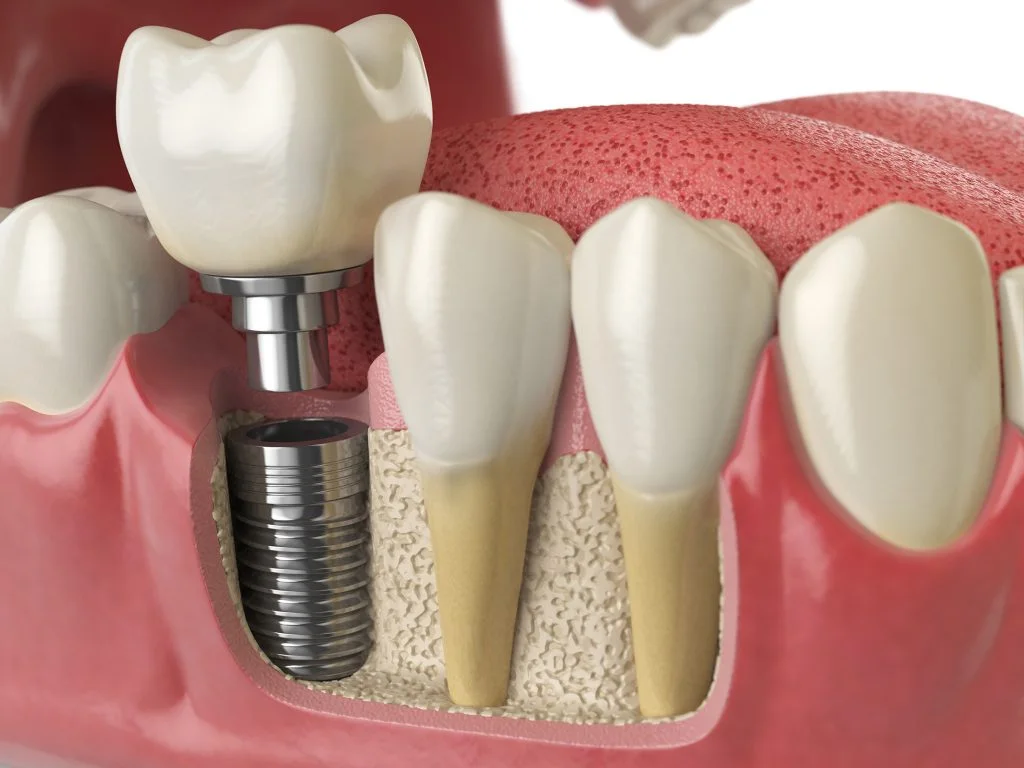
Level 3 • Implants & Surgery
Soft Tissue Grafting: Principles & Techniques
Advanced course on soft tissue grafting principles and techniques around teeth and implants.
- Self-Paced
- 5–6 CE Credits
- 6–8 Hours
- On-Demand
$449View Course

Bone Grafting & Site Development • Clinical Guide
Discover effective soft tissue grafting techniques for dental implants to improve stability and aesthetics, ensuring optimal long-term results.
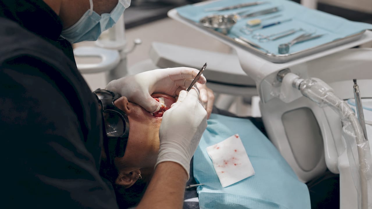
Graft around implants when keratinized tissue width (KTW) falls at or below 2 mm, or when mucosal thickness is insufficient for biological stability and esthetic outcomes. For mucosal thickness augmentation, the subepithelial connective tissue graft (SCTG) is the gold standard. For KTW gain, an apically positioned flap combined with a free gingival graft (APF+FGG) produces the most reliable increases. Xenogeneic collagen matrices (XCM) and acellular dermal matrices (ADM) are reasonable when reducing donor-site morbidity is a priority, but expect less predictable long-term volume stability.
Pro Tip: Before any implant procedure, record baseline KTW and mucosal thickness with a calibrated probe and stent. You cannot track what you did not measure.
Soft tissue grafting around implants is indicated when KTW falls at or below 2 mm or mucosal thickness drops below approximately 2 mm, with SCTG as the gold standard for thickness gain and APF+FGG as the most reliable approach for KTW augmentation.
| Point | Details |
|---|---|
| KTW and MT thresholds | Graft when KTW ≤ 2 mm or MT < 2 mm; both thresholds are linked to marginal bone loss and hygiene compromise. |
| Material hierarchy | SCTG produces 0.8–2.5 mm thickness gains; APF+FGG yields 1.4–3.3 mm KTW gains; XCM/ADM reduce morbidity but produce smaller, less stable gains. |
| Plan for shrinkage | SCTG shrinkage of 0.2–1.1 mm is expected within 6–12 months; augment slightly beyond the minimum target when long-term stability is required. |
| Timing is secondary to technique | Technique and material choice drive outcomes more than timing, but second-stage surgery is a practical and evidence-supported grafting window. |
| Onewd training pathway | Onewd’s hands-on implant and soft tissue grafting courses provide supervised live-patient experience to translate this evidence into predictable clinical outcomes. |
Peri-implant soft tissue deficiencies fall into two distinct clinical problems, and conflating them leads to the wrong treatment plan. The first is inadequate keratinized tissue width, which compromises oral hygiene access and long-term mucosal stability. The second is insufficient mucosal thickness, which predisposes the site to marginal bone loss and esthetic failure regardless of how much keratinized tissue is present.
The Osteology Foundation Consensus Report identifies KTW ≤ 2 mm as the commonly used clinical threshold for augmentation. Sites below this mark tend to accumulate more plaque, bleed more readily on probing, and show higher rates of peri-implant mucositis. Patients with thin, mobile mucosa also report greater discomfort during brushing, which compounds hygiene compliance problems over time.
Measuring KTW is straightforward: use a periodontal probe with a fixed reference stent to record the distance from the mucosal margin to the mucogingival junction. Reproducibility improves when the same clinician uses the same stent at each recall. For anterior sites where esthetics drive the decision, CBCT-to-soft-tissue overlay or transgingival probing with a caliper can add volumetric context, though probe-based measurement remains the clinical standard.
A mucosal thickness below approximately 2 mm is associated with greater marginal bone resorption in multiple systematic reviews. The mechanism is straightforward: thin mucosa cannot buffer the biologic width demands of the implant-abutment interface, and the body compensates by resorbing crestal bone. Meta-analytic data confirm that connective tissue grafting for mucosal thickness is associated with significantly less marginal bone loss compared to non-grafted controls.
Transgingival probing under local anesthesia gives the most direct thickness measurement. Ultrasonic devices and CBCT-based soft-tissue proxies are emerging but not yet standard in most practices.
| Phenotype | Location | Patient Priority | Recommended Action |
|---|---|---|---|
| Thin mucosa, KTW ≤ 2 mm | Anterior esthetic zone | Esthetics + stability | Stage SCTG before or at implant placement |
| Thin mucosa, KTW ≤ 2 mm | Posterior functional zone | Hygiene access | APF+FGG at second-stage or before placement |
| Adequate KTW, thin mucosa | Anterior esthetic zone | Esthetics | SCTG bilaminar approach; consider timing with provisionalization |
| Adequate KTW, adequate mucosa | Any | Maintenance | No graft; monitor at recall |
| Thin mucosa, limited palatal tissue | Any | Reduced morbidity | XCM or ADM with informed consent on predictability limits |
When soft-tissue deficiency is severe enough to compromise primary implant stability or flap closure, staging the graft before implant placement is the safer path. Attempting simultaneous bone and soft-tissue augmentation at a site with minimal attached mucosa increases the risk of membrane exposure and graft failure.
Each technique in the oral soft tissue grafting toolkit has a distinct indication, and none of them is universally superior across all clinical goals.
Harvested from the palate via a partial-thickness trap-door or single-incision technique, the SCTG is placed beneath a split-thickness or coronally advanced buccal flap. It is the gold standard for mucosal thickness augmentation, producing thickness gains of approximately 0.8–2.5 mm at 6–12 months across RCTs. Color match is excellent because the graft is covered by native epithelium. The trade-off is a palatal donor site, which is the primary source of postoperative morbidity.
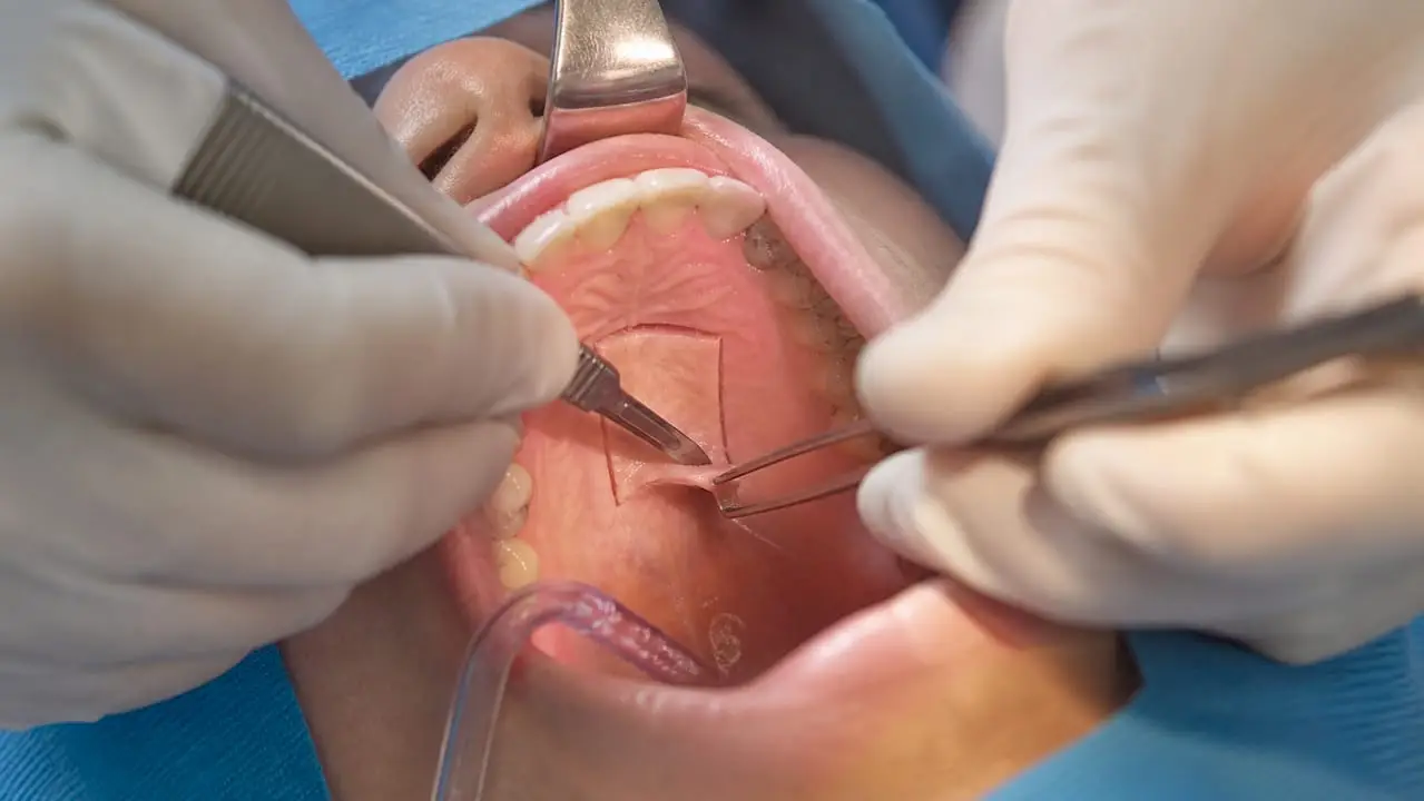
Pros: Best long-term volume stability; superior esthetic integration; well-documented marginal bone preservation.
Cons: Two surgical sites; longer operative time; palatal healing discomfort for 2–4 weeks.
The FGG is an epithelialized graft, also harvested from the palate, placed directly on a periosteal bed. It is most effective for increasing KTW, particularly in posterior sites where color match is less critical. Used with an apically positioned flap, it produces the largest KTW gains in the literature.
Pros: Technically straightforward; reliable KTW increase; durable long-term.
Cons: Color mismatch (pale, opaque appearance); not suitable for anterior esthetic zones; donor-site morbidity similar to SCTG.
The APF repositions existing mucosa apically to expose periosteum and create a new zone of attached tissue. Used alone, it increases KTW but does not add volume. Combined with FGG or SCTG, it becomes the most effective approach for KTW gain. A systematic review reports APF+FGG or APF+SCTG producing KTW gains of approximately 1.4–3.3 mm, outperforming APF alone or no treatment.
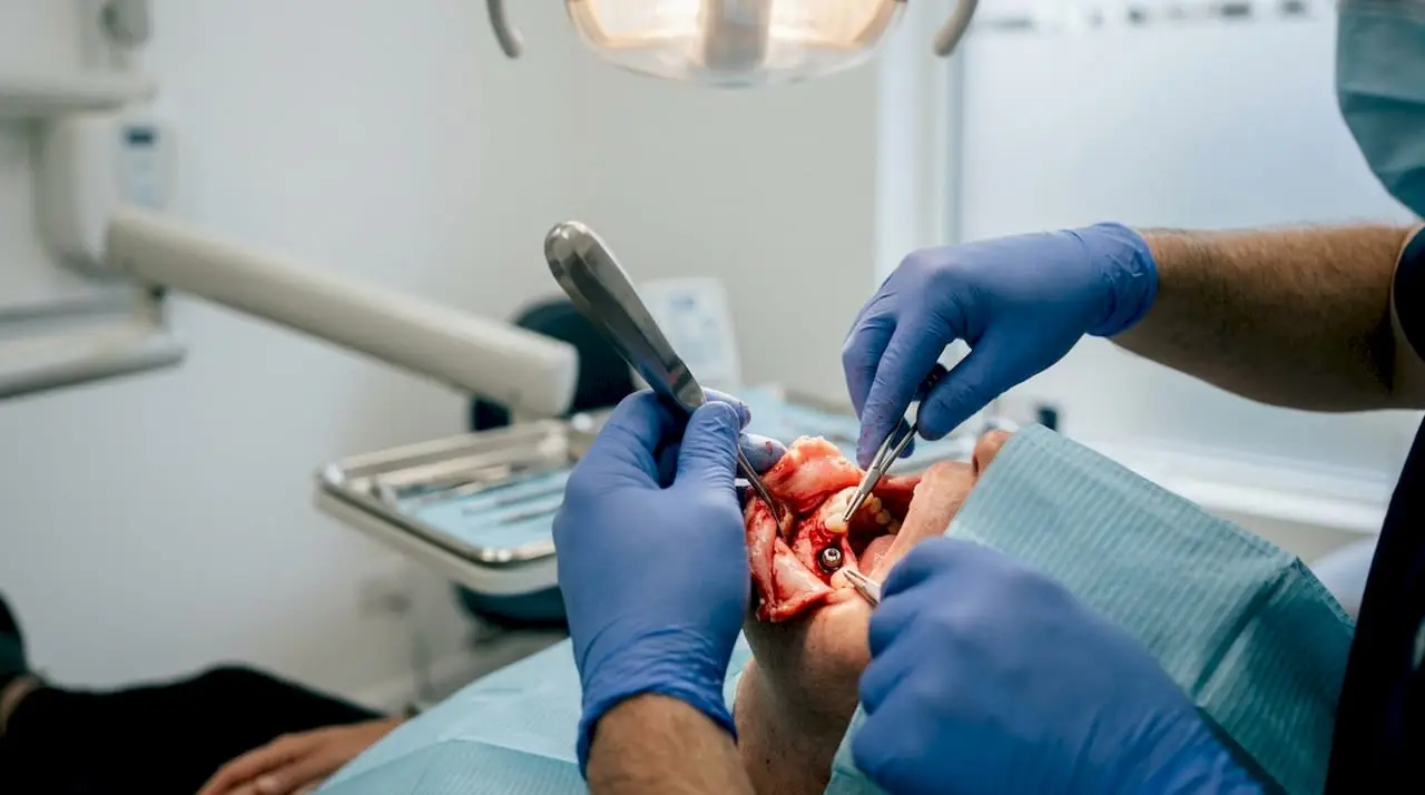
Vestibuloplasty is essentially an APF extended across a broader edentulous span, often used in full-arch implant cases where the vestibular depth is inadequate.
Bilaminar techniques combine a CTG with a coronally advanced flap (CAF) or split-thickness flap. These are preferred in anterior esthetic sites where root or implant surface coverage and volume augmentation are both needed. The two-layer design protects graft vascularity while allowing coronal advancement without tension.
Products like Mucograft (Geistlich) are porcine-derived collagen matrices used as autograft substitutes. They eliminate the palatal donor site entirely, reducing surgery time and patient morbidity. RCT data consistently show lower morbidity scores with XCM compared to SCTG, but gains in KTW and mucosal thickness are smaller and more variable. Long-term volume stability is less predictable.
Pros: No donor site; shorter surgery; lower early morbidity.
Cons: Less predictable volume gain; higher shrinkage variability; greater cost per unit.
ADM products (such as AlloDerm) are human-derived processed dermis. They behave similarly to XCM in clinical outcomes: reduced morbidity versus autograft, but generally inferior long-term thickness gains. ADM may be preferred when palatal tissue is genuinely insufficient or when the patient declines a second surgical site.
Implant hardware complicates flap advancement and suturing. When the healing abutment or provisional crown is present, use interrupted periosteal sutures to anchor the graft apically and sling sutures around the abutment to prevent coronal displacement. Avoid placing sutures directly over the graft center, where tension can compromise vascularity.
Pro Tip: For SCTG around implants, a horizontal mattress suture placed 3–4 mm from the graft margin, combined with a periosteal anchor suture at the base, provides the most reliable stabilization without compressing the graft’s blood supply.
Timing is often treated as a secondary decision, but it directly affects graft stabilization feasibility, the number of surgical entries, and patient morbidity.
| Phenotype | Procedure Complexity | Recommended Timing |
|---|---|---|
| Thin mucosa, adequate KTW | Immediate implant, no bone graft | Simultaneous SCTG at placement |
| KTW ≤ 2 mm, thin mucosa | Delayed implant, staged site | Graft before implant placement |
| KTW ≤ 2 mm, adequate mucosa | Standard delayed implant | APF+FGG at second-stage |
| Any deficiency, missed earlier | Restored implant | Delayed graft with modified flap design |
| Thin mucosa, bone graft needed | Complex simultaneous GBR | Stage graft before or after GBR; avoid simultaneous |
The evidence from a 2026 network meta-analysis suggests that technique and material choice have a greater influence on outcomes than timing alone, but individualized timing decisions based on anatomy and stabilization feasibility remain clinically important.
The literature on soft tissue grafting around implants has grown substantially, though heterogeneity in study design, follow-up duration, and measurement methods limits direct comparisons.
| Source | Intervention | Outcome | Result | Evidence Notes |
|---|---|---|---|---|
| Zucchelli et al. / JCPE systematic review | APF+FGG vs. APF alone | KTW gain | 1.4–3.3 mm with APF+FGG | Moderate heterogeneity; short follow-ups in some studies |
| Osteology Consensus (CLR 2019) | Autogenous grafting (KTW) | Plaque/bleeding indices | Significant improvement vs. control | Consensus-level; RCT base |
| Thoma et al. meta-analysis (CLR) | CTG for mucosal thickness | Marginal bone level | Significantly less bone loss vs. no graft | Moderate evidence; varied follow-up |
| Cafasso et al. NMA 2026 | APF+FGG vs. CTG vs. XCM | KTW and MT | APF+FGG best KTW; CTG best MT; XCM lower gains | Network meta-analysis; largest synthesis to date |
| SCTG vs. XCM RCTs (pooled) | SCTG vs. collagen matrix | MT gain | SCTG: 0.8–2.5 mm; XCM: 0.3–1.2 mm | Lower morbidity with XCM; less gain |
A thickness gain of at least 1 mm is generally considered clinically meaningful for marginal bone preservation. Meta-analytic data confirm that even modest thickness increases reduce marginal bone loss and improve esthetic outcomes across multiple studies. The pink esthetic score (PES) improves most reliably with SCTG in anterior sites, where papilla fill and mucosal contour are the dominant esthetic parameters.
For KTW, the systematic review evidence shows APF+autogenous grafts outperform APF alone or no treatment by a clinically significant margin. Sites treated with APF alone gain some KTW but less reliably, and without the volume benefit.
SCTG shrinkage of approximately 0.2–1.1 mm in thickness (or roughly 10–40 mm³ volumetrically) occurs within 6–12 months of placement. This is not failure; it is expected remodeling. The practical implication: target slightly greater initial augmentation than the minimum threshold when long-term stability is the priority. XCM shrinkage is variable and in some RCTs exceeds that of SCTG in proportional terms, which partly explains the lower net gains at follow-up.
Short follow-up periods (often 3–6 months) dominate the literature, and few studies track outcomes beyond 12 months. Measurement methods vary widely, from probe-based KTW to volumetric scanning, making pooled analyses imprecise. The 2026 network meta-analysis by Cafasso et al. is the most comprehensive synthesis available, but the authors note residual heterogeneity and call for standardized outcome reporting in future trials. Interpret numeric ranges as guidance, not guarantees.
Technical execution determines whether the evidence-based gains you expect actually materialize in your patient.
Partial-thickness palatal flap harvest with careful de-epithelialization of the recipient bed improves graft revascularization. On the buccal side, combining 2–3 mm of full-thickness release with split-thickness advancement reduces tension while preserving perfusion. These two principles together account for most of the difference between a graft that integrates and one that partially necroses.
When implant hardware is present, the healing abutment height affects flap advancement. A shorter healing abutment facilitates coronal advancement; a tall abutment may require a custom provisional to support the graft margin.
The palate between the canine and first molar is the primary harvest zone. Avoid harvesting within 2 mm of the gingival margin to preserve palatal gingival integrity. When palatal tissue is thin (less than 3 mm total thickness including epithelium), consider a single-incision technique rather than a trap-door to reduce the risk of palatal artery injury. If palatal tissue is genuinely insufficient, XCM or ADM becomes the practical alternative rather than a preference.
Pro Tip: A pre-operative CBCT or ultrasonic measurement of palatal tissue thickness at the harvest site takes under two minutes and prevents the unpleasant intraoperative discovery of a thin palate. Target a harvest zone with at least 3.5 mm total tissue thickness.
Complications after soft tissue grafting around implants are manageable when anticipated, but they require a systematic approach rather than reactive improvisation.
Initial epithelialization of the graft surface takes approximately two weeks. During this phase, the graft appears pale and slightly raised; this is normal. Progressive integration and vascular ingrowth continue through 4–12 weeks, during which the graft softens and begins to match surrounding tissue color. Final volumetric stability is not reached until 6–12 months, which is why outcome measurements taken at 3 months underestimate shrinkage.
Work through these steps in sequence for every implant patient with a potential soft-tissue deficiency.
Pro Tip: Photograph the site with a calibrated ruler at baseline, immediately post-op, and at 3 and 6 months. Volumetric photo documentation is the fastest way to identify early shrinkage before it becomes a clinical problem.
Soft tissue grafting around implants is a technique-sensitive procedure. Reading the evidence is necessary but not sufficient; the gap between knowing the algorithm and executing it predictably closes only through structured, progressive hands-on training.
Refer to a periodontist or oral surgeon when the case involves a failed previous graft, active peri-implant disease requiring concurrent treatment, a severely limited tissue envelope (less than 1 mm attached mucosa with no vestibular depth), or a complex anterior esthetic case where a single graft failure would compromise the final restoration. Recognizing the limits of your current skill level is a clinical competency, not a shortcoming.
For clinicians building this skill set, hands-on implant training that integrates soft tissue management into the implant placement workflow is the most efficient path to predictable outcomes. The implants and surgery course catalog at Onewd covers the full procedural arc from site development through soft-tissue management.
The literature on soft tissue grafting around implants is more consistent than it was a decade ago. APF+FGG for KTW, SCTG for mucosal thickness, XCM when morbidity is the constraint: the hierarchy is reasonably clear. What the systematic reviews cannot tell you is how to manage the moment when your flap advancement is 1 mm short of tension-free closure, or when the palatal harvest comes out thinner than planned.
The clinicians who produce the most predictable outcomes are not necessarily those who have read the most meta-analyses. They are the ones who have done enough supervised cases to have internalized the tactile feedback of a well-placed periosteal suture and the visual cue of a graft that is adequately vascularized versus one that is not. Evidence sets the target; technique gets you there.
There is also a tendency in the field to treat material selection as the primary decision and technique as secondary. The data suggest the opposite: a well-executed SCTG with proper flap design outperforms a poorly executed XCM placement every time, even accounting for the morbidity difference. Invest in the technique first, then optimize the material choice.
Knowing the indications and the algorithm is the starting point. Executing a bilaminar SCTG or an APF+FGG predictably in a live patient requires a different kind of preparation, and that is exactly where structured, mentorship-driven training pays off.
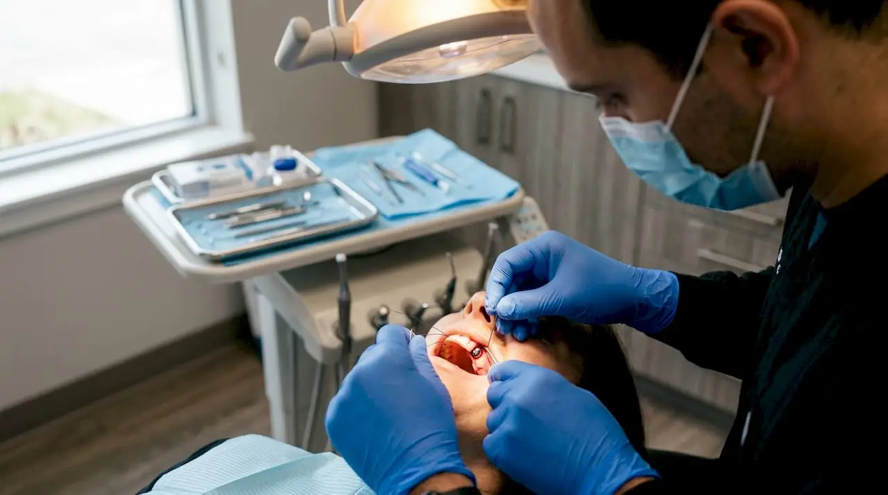
Onewd’s hands-on implant training programs are built around the principle that supervised repetition, not passive observation, is what converts clinical knowledge into reliable outcomes. The soft tissue grafting course covers flap design, CTG harvest, graft stabilization around implant hardware, and suturing sequences, with live-patient modules that put you in the operator’s seat under direct mentorship. Courses are available in both online and in-person formats, with CE credits upon completion.
If you are ready to move from reading the evidence to applying it, browse the full training course catalog and find the format that fits your schedule and current skill level.
Educational disclaimer: Content is provided for professional education and is not a substitute for a clinician's independent judgment, applicable standards, manufacturer instructions, or relevant laws and regulations.
Related Training
Level 3 • Implants & Surgery
Advanced course on soft tissue grafting principles and techniques around teeth and implants.
Level 1 • Implants & Surgery
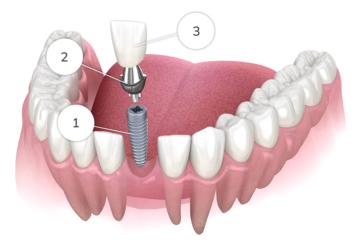
Self-paced online lecture series on simple single-tooth implant placement in the anterior and posterior.
Bone Grafting • Clinical Guide
Discover how soft tissue grafting around implants can enhance mucosal health. Learn effective techniques to improve implant outcomes.
17 min read · Updated August 27, 2026
Bone Grafting • Comparison
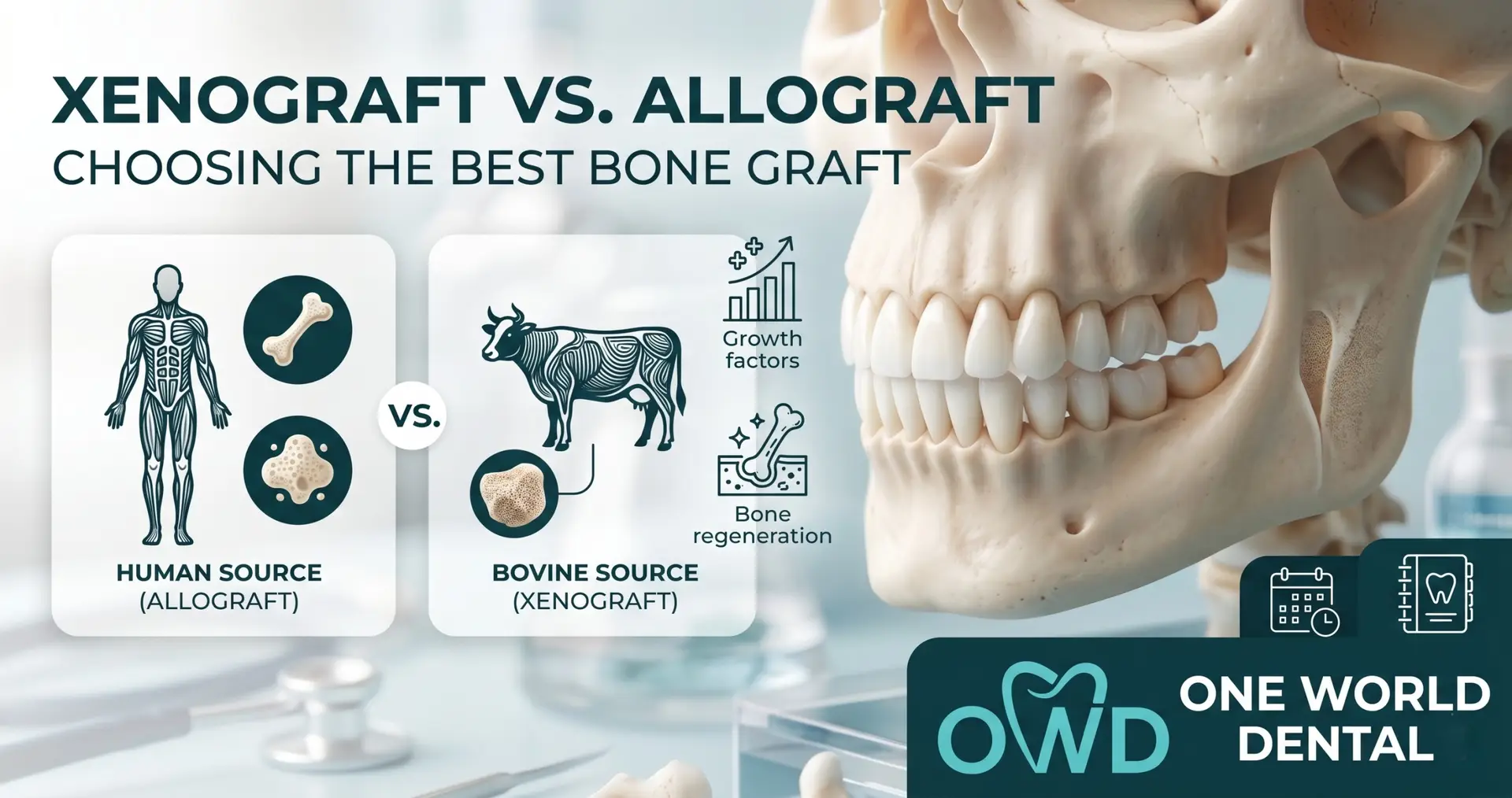
Master bone graft selection. Contrast resorption rates, remodeling timelines, and clinical indications for allografts and xenografts.
6 min read · Updated May 19, 2026
Bone Grafting • Procedure Guide
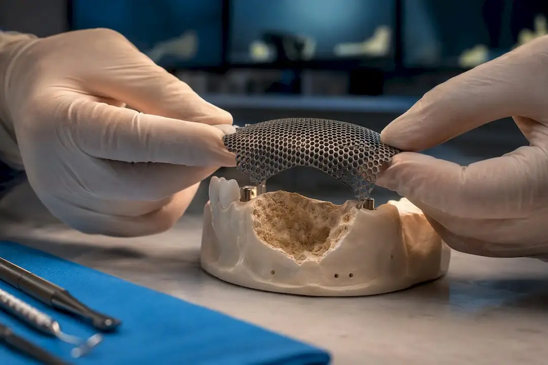
For clinicians: a defect-based ridge augmentation guide that matches technique to anatomy, explains mesh and CAD/CAM workflows, plans 4–6 month staging,...
14 min read · Updated September 5, 2026