In modern implantology, selecting a bone grafting material is no longer about simply filling a void. Predictable hard-tissue regeneration requires an understanding of cellular biology, host tissue responses, and the specific degradation timelines of your biomaterials. Matching the wrong graft material to your clinical objective is a leading cause of premature ridge collapse and implant failure.
At One World Dental, our advanced surgical and implant training modules focus heavily on evidence-based biomaterial selection. We move clinicians past the marketing fluff and dive deep into the micro-architecture of bone grafts.
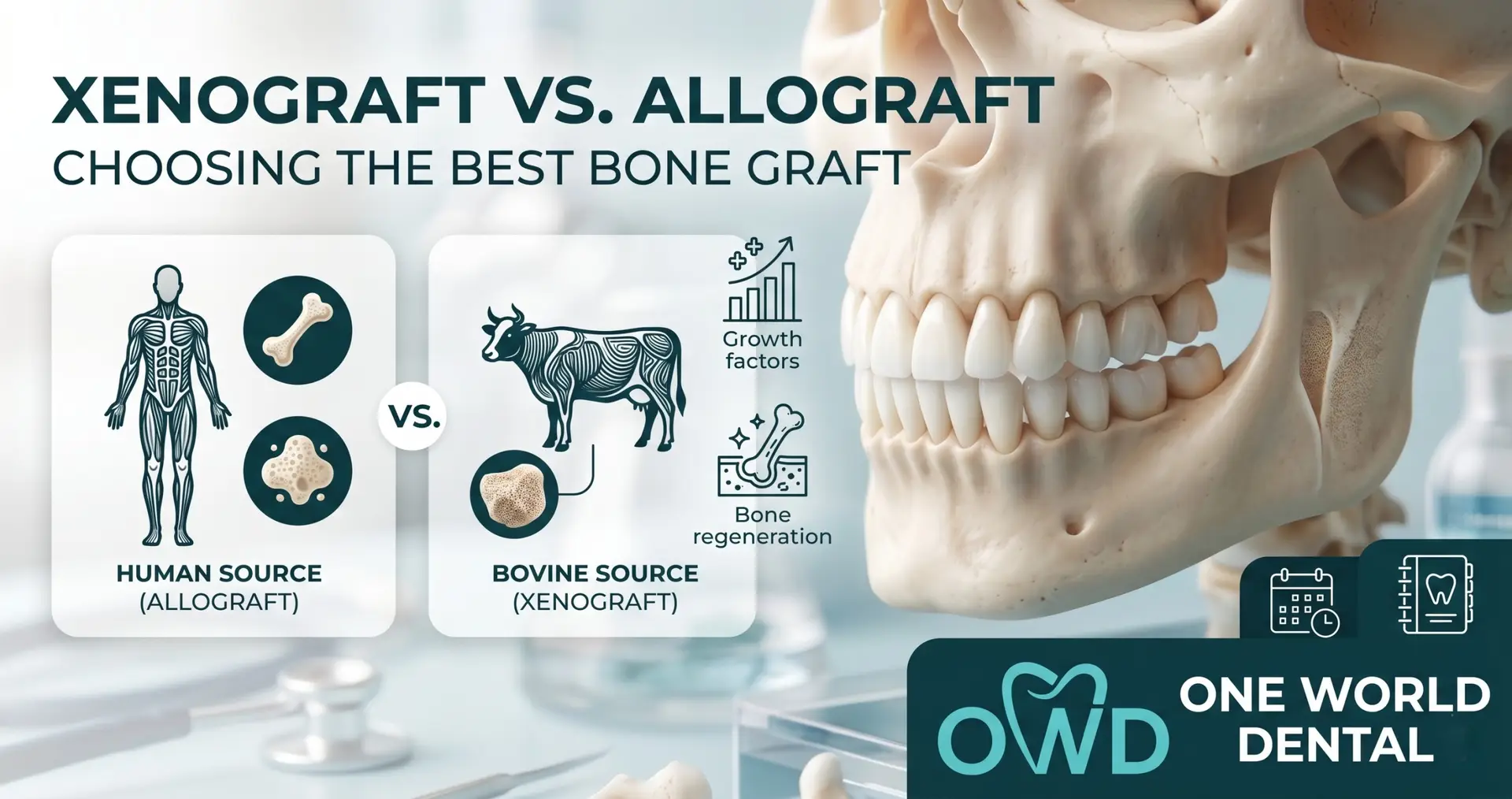
Below, we analyze the critical differences between allografts and xenografts, their biological mechanisms, and how to select the ideal material based on your clinical workflow.
The Biological Difference: Osteoconduction vs. Osteoinduction
To make an informed clinical choice, you must first differentiate how these materials interact with the host healing bed:
Osteoinduction (The Signal): The material actively recruits and stimulates undifferentiated mesenchymal stem cells from the host bed, inducing them to differentiate into bone-forming osteoblasts. This process is heavily driven by native bone morphogenetic proteins (BMPs).
Osteoconduction (The Scaffold): The material acts as a passive, physical matrix through which local blood vessels and osteoprogenitor cells can migrate. It maintains space but does not actively stimulate bone formation.
Allografts: Rapid Remodeling and Vital Bone Formation
Derived from human donor tissue (processed via strict tissue-bank protocols), allografts are the workhorses of standard socket preservation and localized ridge augmentations.
Types of Allograft
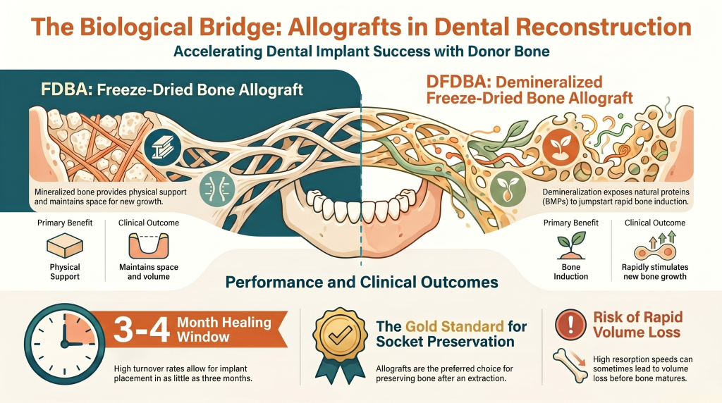
Mineralized Freeze-Dried Bone Allograft (FDBA): Provides excellent osteoconductive space maintenance while retaining structural strength. Remodels predictably into vital host bone over a moderate timeline.
Demineralized Freeze-Dried Bone Allograft (DFDBA): Acid extraction removes the mineral content, exposing the underlying collagen matrix and native BMPs. This shifts the material’s property toward being primarily osteoinductive, accelerating early osteogenesis.
Clinical Remodeling Timeline
Allografts turn over rapidly. Depending on whether you choose a cortical, cancellous, or blended preparation, you can generally expect predictable host-bone replacement and high percentages of vital bone within 3 to 4 months, making it ideal for standard implant staging.
Derived from a non-human species—most commonly bovine (cow) or porcine (pig) origins—xenografts undergo extreme thermal and chemical processing to completely strip all organic material, leaving behind a pure, sterile hydroxyapatite mineral matrix.
The Mechanism of Action
Xenografts are strictly osteoconductive. Because the processing eliminates all proteins and growth factors, the material provides zero osteoinductive signaling.
Clinical Remodeling Timeline
The hallmark feature of a xenograft is its incredibly slow resorption rate. The crystalline structure of bovine bone resists osteoclastic breakdown. Years after placement, histological sections will still show xenograft particles encapsulated by new vital host bone. This makes xenografts the gold standard for long-term volume maintenance, particularly when resisting the heavy resorptive forces of the buccal plate in the aesthetic zone.
Clinical Decision: Mapping Materials to Indications
When deciding between these two biomaterials chairside, use this clinical matrix as a reference guide:
Clinical Indication
Preferred Material
Primary Clinical Rationale
Aesthetic Zone Socket Preservation
Xenograft (or Allograft/Xenograft Blend)
Prevents the inevitable 40–60% structural collapse of the delicate buccal plate over time due to its slow resorption rate.
Posterior Socket Preservation (Early Staging)
Allograft (FDBA/DFDBA)
Rapidly remodels into dense, vital host bone, allowing predictable implant placement within 12 to 16 weeks.
Maxillary Sinus Floor Elevation
Allograft/Xenograft Blend (50/50)
The allograft drives early vital bone formation near the native sinus walls, while the xenograft maintains the critical sub-antral volume against sinus pneumatization.
Horizontal Ridge Augmentation (GBR)
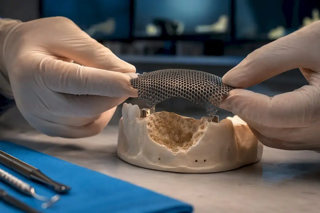
Mineralized Cortical Allograft + Membrane
Provides the rigid, high-turnover scaffolding required to bulk out a deficient ridge before or during implant fixture placement.
Advanced Protocols: Biologics and “Sticky Bone” Hydration
In our advanced One World Dental surgical masterclasses, we teach clinicians how to radically upgrade the handling and biological potential of both allografts and xenografts using autologous blood concentrates.
By utilizing localized venipuncture and specific centrifugation protocols, you can generate Liquid PRF (i-PRF). Hydrating your particulate allograft or xenograft with i-PRF creates a cohesive, moldable “sticky bone” matrix.
This protocol offers two massive clinical advantages:
Mechanical Stability: Completely eliminates the frustration of particulate graft washing out or migrating under the flap.
Biological Acceleration: Infuses a strictly osteoconductive matrix (like a xenograft) with a high concentration of autologous growth factors, leukocytes, and a fibrin network, turning a passive scaffold into a highly active healing environment.
Mastering Biomaterial Mechanics for Clinical Predictability
Achieving long-term implant success requires moving past a “one-size-fits-all” approach to bone grafting. Understanding the precise cellular interactions, structural limits, and resorption curves of your biomaterials is what separates guess-work from true clinical mastery.
At One World Dental, our intensive, hands-on surgical training courses are designed to give you the confidence to execute advanced Guided Bone Regeneration (GBR), sinus lifts, and sinus membrane management protocols with predictable, evidence-based results.
Why not use 100% xenograft for all bone grafting procedures if it maintains volume so well?
While volume maintenance is highly desirable, a successful implant requires a specific percentage of living, vascularized vital host bone to undergo osseointegration. Because xenografts resorb at an incredibly slow rate, utilizing a 100% xenograft in a large, contained defect can result in a high concentration of unresorbed graft particles and a lower percentage of vital bone at your implant site, potentially compromising primary and secondary stability.
What is the clinical benefit of blending cortical and cancellous allograft together?
Blending combines two distinct remodeling profiles. Cancellous allograft bone has a highly porous micro-architecture that allows rapid vascularization and swift cellular turnover (fast bone formation). Cortical allograft bone is dense and takes significantly longer to resorb, providing structural rigidity and space maintenance during the early phases of healing. A standard 50/50 blend gives you the ideal balance of early stability and rapid vital bone remodeling.
How do I handle a patient who objects to the use of xenograft or allograft due to religious or ethical reasons?
Pre-operative informed consent must always include a clear discussion on the origin of your biomaterials. If a patient declines human-derived (allograft) or animal-derived (xenograft) tissue due to dietary, ethical, or religious practices, you must pivot to a synthetic/alloplastic material (such as beta-tricalcium phosphate or bioactive glass). Our training modules include specific communication frameworks to help your team navigate these treatment modifications seamlessly.
When performing an immediate implant placement, should I graft the “jumping distance” with an allograft or a xenograft?
A xenograft is highly preferred for grafting the horizontal gap (jumping distance) between the implant body and the intact buccal plate. Because the buccal plate in an immediate site is incredibly prone to osteoclastic resorption and remodeling, placing a slow-resorbing xenograft particulate directly against the internal surface of the buccal wall acts as a long-term shield, preventing post-operative soft tissue recession and preserving the aesthetic profile.
Does the hydration protocol of the bone particulate actually impact the surgical outcome?
Yes. Particulate bone should never be placed dry into a surgical bed. Hydrating the graft with sterile saline, sterile water, or ideally, autologous blood concentrates (like PRF) activates the surface chemistry of the particles, facilitating early protein adsorption and cellular adhesion. Furthermore, over-saturating or compressing the graft too tightly into the socket can occlude the micro-scaffolding, preventing angiogenesis and leading to graft necrosis rather than regeneration.
Can I place an implant directly into a site previously grafted with DFDBA sooner than an FDBA site?
Theoretically, yes. Because Demineralized Freeze-Dried Bone Allograft (DFDBA) is primarily osteoinductive, it bypasses the extensive time required for the host to resorb mineralized matrix crystals, often accelerating early bone formation. However, because it lacks mineral density, it provides very little structural stability. If primary implant stability relies heavily on the graft site’s mechanical strength, a mineralized FDBA or a cortical blend is required, which requires a slightly longer maturation window before fixture placement.
Educational disclaimer: Content is provided for professional education and is not a substitute for a clinician's independent judgment, applicable standards, manufacturer instructions, or relevant laws and regulations.
For clinicians: a defect-based ridge augmentation guide that matches technique to anatomy, explains mesh and CAD/CAM workflows, plans 4–6 month staging,...