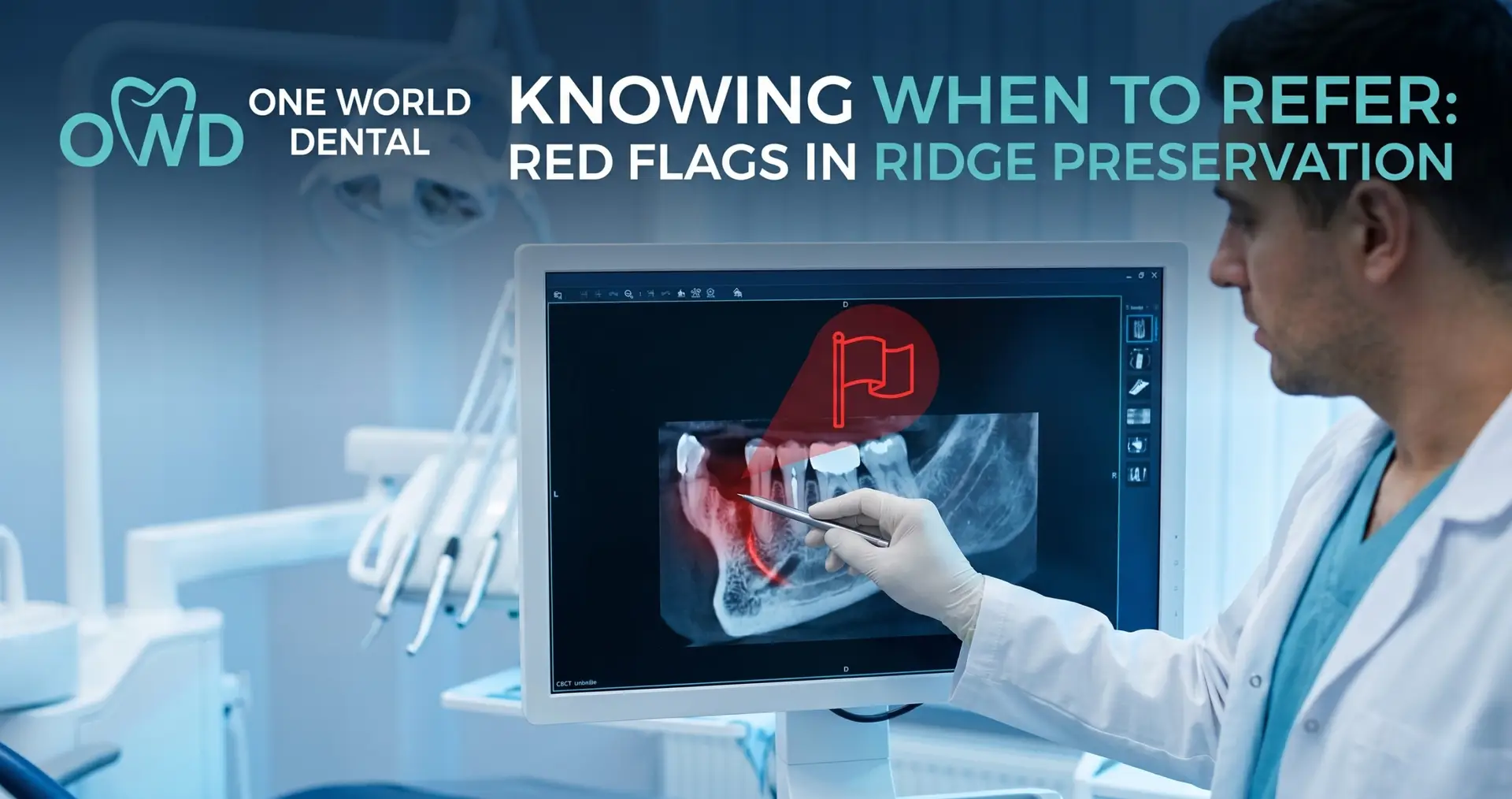
Knowing when to refer: red flags in ridge preservation is a critical clinical competency for the general practitioner managing the transition from extraction to implant placement. While alveolar ridge preservation (ARP) is often viewed as a routine procedural adjunct, the biological complexity of the alveolar housing necessitates a sophisticated understanding of when a case exceeds the scope of standard primary care. One World Dental serves as a specialized resource for clinicians encountering these complex biological and anatomical hurdles.
The Goal of Alveolar Ridge Preservation (ARP)
The primary objective of ARP is the attenuation of the physiological ridge resorption that follows tooth extraction. Following the luxation of a tooth, the bundle bone—which is tooth-dependent—undergoes inevitable catabolism.
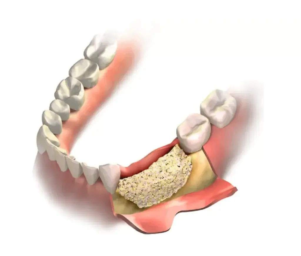
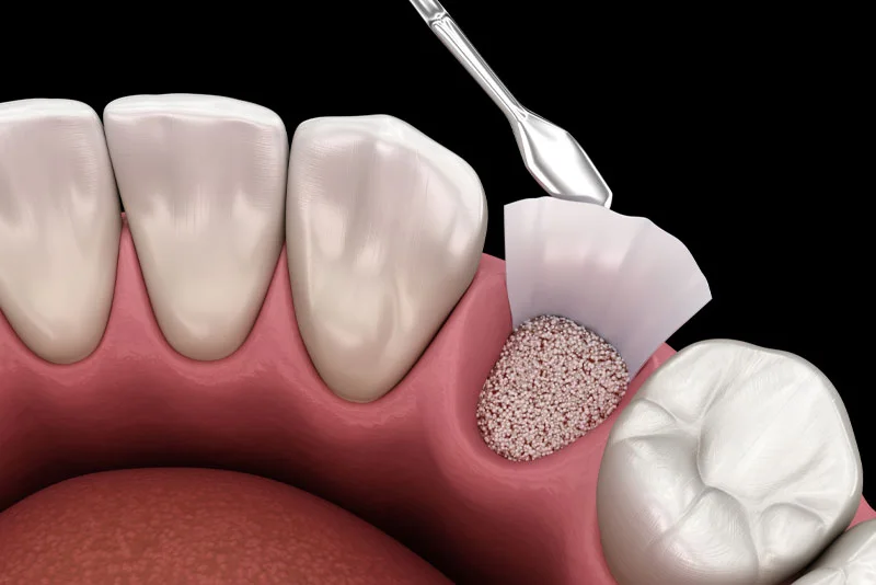
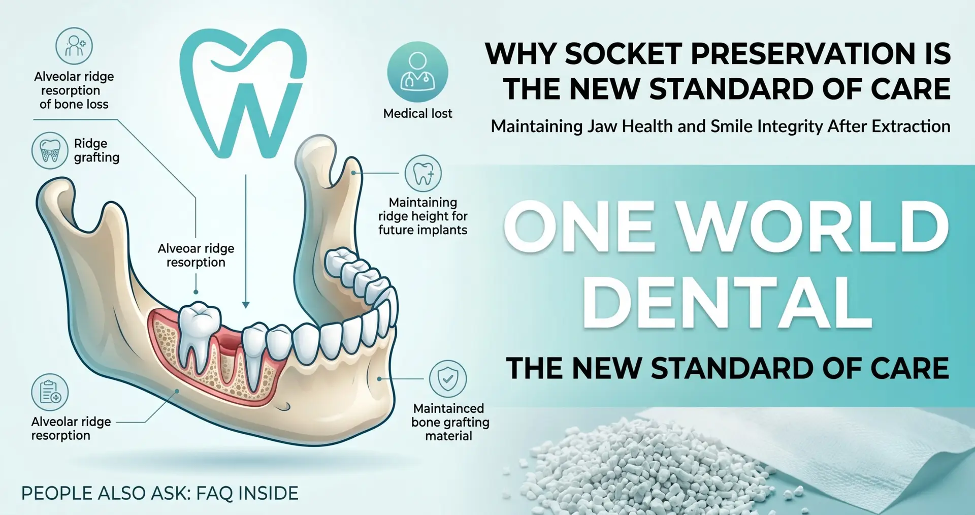
Within the first six months post-extraction, the horizontal dimension of the ridge can decrease by up to 50%. ARP utilizes osteoconductive or osteoinductive scaffolds to maintain the “envelope” of the hard tissue. However, practitioners must distinguish between a simple socket fill and a complex ridge reconstruction. When the clinical objective shifts from preserving existing bone to regenerating lost buccal plates, a referral to One World Dental ensures that advanced grafting techniques are employed to maintain the long-term restorative platform.
Pre-Surgical Red Flags: When to Refer Before the Extraction
Predictable outcomes in ridge preservation are often determined before the first periotome is utilized. A thorough systemic and local assessment is the first line of defense against graft failure.
Medical Contraindications and Host Response
A patient’s systemic health directly dictates the success of the angiogenesis required for graft integration. Clinicians should screen for the following “red flags”:
Metabolic Dysregulation: Patients with an HbA1c exceeding 7.0% exhibit impaired polymorphonuclear leukocyte function and collagen synthesis. These patients are at a higher risk for secondary graft infection and marginal bone loss.
Antiresorptive Therapy: A history of IV bisphosphonates (e.g., Zometa) or high-dose oral bisphosphonates presents a significant risk for Medication-Related Osteonecrosis of the Jaws (MRONJ). If the systemic risk is high, surgical intervention should be managed by a specialist.
Vascular Impairment: Heavy smoking (defined as >10 cigarettes per day) creates a localized hypoxic environment. This inhibits the migration of osteoblasts to the graft site, often resulting in “dry” non-integrated granules.
Anatomical Complexity and CBCT Analysis
Pre-operative imaging, specifically Cone Beam Computed Tomography (CBCT), is essential for identifying “no-go” zones.
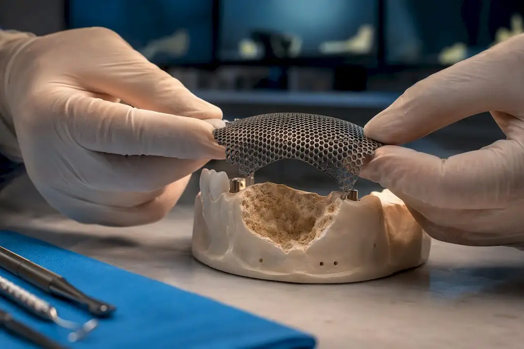
Type III Sockets: If the buccal plate is missing more than 50% of its vertical height, standard ARP protocols are insufficient. These cases require Guided Bone Regeneration (GBR) with reinforced membranes, a procedure with a steeper learning curve.
Neurological Proximity: When the root apices are in direct contact with the Inferior Alveolar Nerve (IAN) canal, the risk of paresthesia during debridement or graft packing increases.
Maxillary Sinus Pneumatization: If there is less than 2mm of bone between the root apex and the sinus floor, the risk of an Oroantral Communication (OAC) is high. One World Dental specializes in simultaneous sinus floor elevation and grafting to mitigate these risks.
Intra-Operative “Stop Signs” for the General Dentist
Even with a planned extraction, the clinical reality can change mid-procedure. Recognizing these intra-operative red flags is crucial for risk management.
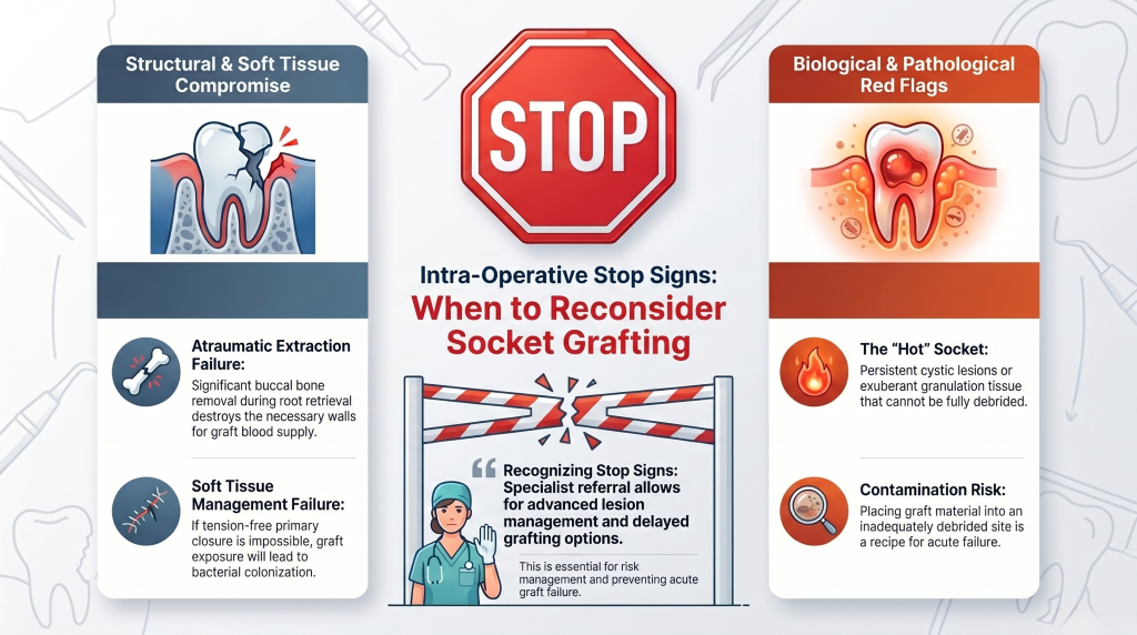
Atraumatic Extraction Failures
The success of ARP is predicated on an atraumatic extraction. If the procedure evolves into a multi-rooted surgical extraction requiring significant buccal bone removal to retrieve root fragments, the “foundation” for the graft is lost. Excessive ostectomy reduces the number of bony walls available to provide blood supply to the graft.
Persistent Pathology
If, upon curettage, the clinician finds a persistent cystic lesion or exuberant granulation tissue that cannot be fully debrided, the site is “hot.” Placing a bone graft into a contaminated or inadequately debrided socket is a recipe for acute graft failure. Specialist referral allows for advanced lesion management and delayed grafting if necessary.
Soft Tissue Management
A frequent cause of graft failure is the inability to achieve primary closure when required. If the clinician cannot mobilize a tension-free flap to cover a non-resorbable membrane or a large graft volume, the site will likely dehisce. Exposure of the graft leads to bacterial colonization and sequestration of the material.
Post-Surgical Red Flags: Identifying Graft Failure
Monitoring the healing phase (0–14 days) is essential. Clinicians must be able to distinguish between normal postoperative inflammation and early-stage graft failure.
Clinical Signs of Infection
Suppuration and Exudate: Any presence of purulent discharge or “creamy” exudate around the incision line after day 3 indicates bacterial infiltration.
Graft Sequestration: While minor “shedding” of granules can occur, the loss of large portions of the graft (sequestration) indicates a lack of clot stability or an active inflammatory rejection.
Persistent Pain: Pain that is non-responsive to NSAIDs or that increases in intensity after the fourth postoperative day is a primary indicator of localized osteitis or graft infection.
Radiographic Red Flags: The 4-Month Scan
The definitive assessment of ARP success occurs at the 4-to-6-month mark via radiographic evaluation.
Failure of Mineralization: On a periapical or CBCT scan, the graft should show increasing radiopacity over time. If the site remains radiolucent or shows a “moth-eaten” appearance, the material has likely been replaced by fibrous connective tissue rather than bone.
The Radiolucent Border: A dark halo surrounding the graft material suggests that the body has encapsulated the granules in fibrous tissue instead of integrating them. This “non-union” will not provide the primary stability needed for an implant.
Significant Vertical Resorption: Despite ARP, some bone loss is expected. However, if the crestal height has dropped significantly below the neighboring cementoenamel junctions (CEJs), a secondary augmentation procedure at One World Dental may be required before implant placement.
The Referral Threshold: 5 Non-Negotiable Signs to Refer
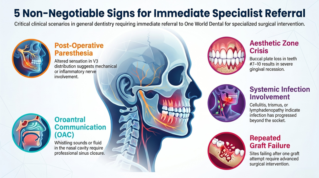
To protect both the patient and the practice, the following scenarios should prompt an immediate referral to One World Dental:
Post-Operative Paresthesia: Any altered sensation in the V3 distribution suggests mechanical or inflammatory nerve involvement.
Oroantral Communication (OAC): If a “whistling” sound is heard or fluids pass into the nasal cavity, the sinus must be professionally closed to prevent chronic sinusitis.
The Aesthetic Zone Crisis: In the maxillary anterior (Teeth #7–10), the buccal plate is often <1mm thick. Any loss of this plate during extraction will result in severe gingival recession. These cases require the expert soft-tissue management provided by a specialist.
Systemic Involvement: If the patient develops facial cellulitis, trismus (limited opening), or lymphadenopathy, the infection has progressed beyond the socket.
Repeated Graft Failure: If a site has failed to heal after one attempt at grafting, the biological “host bed” is compromised and requires advanced surgical intervention.
Clinical Controversies: Flap vs. No-Flap
The “flapless” approach is often marketed as the gold standard for patient comfort. However, it can be a “red flag” for clinicians who cannot visualize the integrity of the socket walls. If you cannot see the defect, you cannot treat it properly. At One World Dental, we utilize a patient-specific approach—employing flapless techniques only when the buccal plate is confirmed intact via 3D imaging, and full-thickness flaps when ridge augmentation is the primary goal.
Conclusion: The Professional Partnership
The decision to refer is not a sign of clinical weakness but a hallmark of professional excellence. By recognizing the red flags in ridge preservation—from systemic contraindications to intra-operative complications—you ensure that your patients receive the most predictable restorative outcomes.
One World Dental is committed to being your partner in complex oral surgery. Whether you need a second opinion on a CBCT scan or a specialized surgeon to handle an aesthetic zone reconstruction, we are here to support your practice.
What is the best graft material for a GP to use in ridge preservation? Most clinicians find allografts (human bone) or xenografts (bovine bone) the most predictable. Allografts tend to turnover faster, while xenografts are excellent at maintaining long-term volume due to their slow resorption rate.
How long should I wait after ARP before placing an implant? Typically, 4 to 6 months is required. This allows for the transition from the graft scaffold to vital, mineralized host bone capable of achieving osseointegration.
Can I place a bone graft if the tooth had an active abscess? Yes, but only after meticulous mechanical debridement and chemical disinfection (e.g., with saline or Chlorhexidine). If the infection is acute and purulent, it is often safer to debride, wait 2-3 weeks, and perform a delayed graft.
What is the most common reason for ridge preservation failure? The most common reasons are bacterial contamination (infection), lack of graft stability (the granules moving too much), and early exposure of the protective membrane.
When is a membrane absolutely necessary? A membrane should be used whenever there is a deficiency in the socket walls (e.g., a missing buccal plate) or when you need to prevent the fast-growing gum tissue from “invading” the slow-growing bone graft space.
Should I refer if I see a sinus perforation during extraction? If the perforation is larger than 2mm, a referral is highly recommended. Special techniques, such as a “figure-eight” suture, collagen plugs, or advanced flap advancement, are necessary to ensure the hole closes permanently.
Educational disclaimer: Requirements differ by state and licensing authority and change over time. Verify current requirements with your licensing board.
For clinicians: a defect-based ridge augmentation guide that matches technique to anatomy, explains mesh and CAD/CAM workflows, plans 4–6 month staging,...