Choosing between a full-thickness and a split-thickness flap is one of the most critical decisions a clinician makes in oral surgery. The wrong tissue reflection technique can lead to unwanted bone loss, poor soft tissue coverage, or flap necrosis. This guide breaks down the clinical indications, anatomical principles, and surgical workflows so you can choose the right flap with complete confidence.
Understanding the Fundamentals: Anatomical & Histological Differences
To pick the correct flap, you first need to understand what tissues are being lifted.
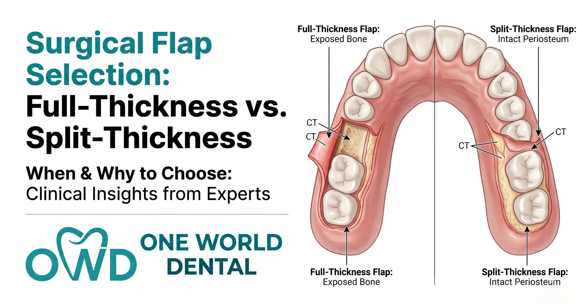
Defining the Full-Thickness (Mucoperiosteal) Flap
A full-thickness flap includes the epithelium, connective tissue, and the periosteum. Using a blunt elevator, you lift all soft tissue clean off the underlying bone. This exposes the bare cortical bone surface.
Defining the Split-Thickness (Partial-Thickness) Flap
A split-thickness flap sharp-dissects through the tissue. It leaves the periosteum and a thin layer of connective tissue attached to the bone. You only lift the epithelium and part of the underlying connective tissue layer.
Vascularity and Blood Supply Mechanics: Mucosa vs. Periosteum
The periosteum carries a dense network of blood vessels. When you lift a full-thickness flap, you temporarily cut off the outer blood supply to the cortical bone.
With a split-thickness flap, the periosteum stays intact on the bone. This protects the bone’s blood supply and provides a vascular bed to nourish soft tissue grafts placed over it.
Key Factors Influencing Flap Selection
Before making your first incision, evaluate these three anatomical factors:
Extent of Bone Access Needed: If you must see, reshape, or place implants directly into bone, you need direct access.
Periodontal Biotype (Thin vs. Thick Tissue): Thin biotypes (under 1.5 mm thick) are fragile. Splitting a thin flap risks tearing or perforating the tissue.
Anatomical Vulnerabilities: Look for bone fenestrations, dehiscences, or thin buccal plates on your CBCT scan. Stripping periosteum off very thin bone can cause irreversible crestal resorption.
Primary Indications for Choosing Full-Thickness Flaps
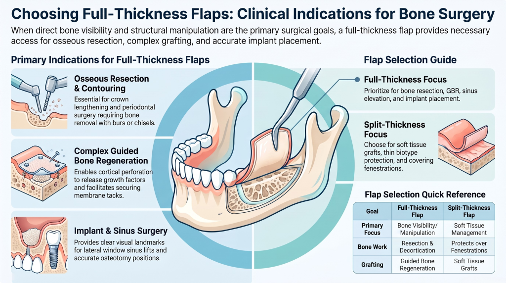
Select a full-thickness flap when direct bone visibility and structural manipulation are your primary goals.
       [ Surgery Requiring Bone Access ]
                      |
        Is direct bone exposure needed?
             /                 \
          (YES)                (NO)
           /                     \
[Full-Thickness Flap]     [Split-Thickness Flap]
 - Bone Resection          - Soft Tissue Grafts
 - Guided Bone Regen.      - Thin Biotype Protection
 - Sinus Elevation         - Apically Positioned Flap
 - Implant Placement       - Over Fenestrations
Osseous Resective Surgery and Direct Bone Manipulation
When performing crown lengthening or resective periodontal surgery, you must remove or reshape bone. Full-thickness exposure allows clear access for burs and chisels.
Complex Guided Bone Regeneration (GBR) and Grafting Procedures
Large bone grafts require full access to the bony bed. Exposing the bone lets you perform cortical perforation (decortication) to release growth factors and secure membrane tacks.
Open Flap Debridement and Extensive Root Planing
Deep intra-bony defects require direct visual contact. Reflecting a full-thickness flap lets you thoroughly clean root surfaces and deep bony pockets.
Multiple Implant Placements and Sinus Lift Elevations
Placing implants, performing lateral window sinus lifts, or expanding ridges require clear visual landmarks to ensure accurate osteotomy positions.
When to Choose Split-Thickness Flaps Instead
Choose a split-thickness flap when your primary goal is soft tissue management or protecting underlying structures.
Mucogingival Procedures and Soft Tissue Grafting: Connective tissue grafts (CTGs) and free gingival grafts (FGGs) need a vascular receiving bed to survive. Leaving the periosteum attached supplies this critical blood flow.
Apically Positioned Flaps Without Exposing Bone: If you move tissue apically to increase keratinized zone width, splitting the flap lets you suture and anchor the tissue directly to the intact periosteum.
Preserving Bone Vascularity in Thin Biotypes: In patients with paper-thin buccal bone, stripping the periosteum can cause the thin bone plate to resorb completely.
Surgeries Over Fenestrations or Dehiscences: If a root is exposed through a bone window, sharp dissection prevents tearing tissue over the bare root.
Comparative Analysis: Healing, Complications, and Recovery
Comparing post-operative responses helps clinicians pick the least invasive option for each scenario.
Clinical Factor
Full-Thickness (Mucoperiosteal) Flap
Split-Thickness (Partial-Thickness) Flap
Primary Surgical Goal
Maximum access to underlying bone
Soft tissue coverage & periosteal preservation
Cortical Bone Response
Temporary ischemia; ~0.5–1 mm crestal bone loss
Preserves periosteal blood supply; minimal bone loss
Surgical Difficulty
Lower (blunt elevation along bone)
Higher (requires precise micro-blade dissection)
Flap Mobility
Limited by periosteal tension
High mobility after releasing periosteal tension
Suturing Anchor
Bone anchor or interdental ties
Direct periosteal anchoring options
Post-Operative Bone Resorption Rates
Stripping periosteum causes temporary bone ischemia. Research shows full-thickness reflection leads to an average of 0.5 to 1.0 mm of marginal bone resorption during healing. Split-thickness flaps prevent this outer bone loss because the periosteal micro-vessels remain intact.
Patient-Reported Post-Operative Pain and Swelling
Full-thickness flaps involve blunt elevation that detaches periosteal fibers from bone, often leading to moderate swelling and aching pain. Split-thickness flaps cause less deep bone aching, though sharp sharp-dissection over a wider surface area can cause temporary superficial discomfort.
Flap Necrosis and Wound Dehiscence Risks
Split-thickness flaps require precise execution. If you make the flap too thin (under 1 mm), you risk cutting off its internal blood supply, leading to partial necrosis or flap tears during suturing. Full-thickness flaps are thicker and less prone to intra-operative tearing.
Step-by-Step Surgical Technique and Equipment Considerations
Proper technique and correct tools are key to clean tissue reflection.
Essential Instruments
Full-Thickness Flaps: Use sharp #15 or #15c scalpel blades for sulcular incisions, followed by periosteal elevators (e.g., Periosteal Elevator #9 or Buser) to lift tissue smoothly.
Split-Thickness Flaps: Use micro-surgical scalpels (e.g., #64 or #69 blades) or sharp #15c blades. Micro-tissue forceps are essential to hold delicate tissue without crushing it.
Incision Protocols and Tissue Reflection Mechanics
For a full-thickness flap, keep your blade pressed firmly against the bone. Once you hit bone, insert the periosteal elevator beneath the periosteum and push firmly against hard tissue to lift the entire complex.
For a split-thickness flap, keep your blade angled parallel to the bone surface. Use light, pulling strokes to split the connective tissue layer, leaving a pink bed of periosteum behind on the bone.
Suturing Techniques and Anchor Strategies
Full-thickness flaps rely on mattress sutures or sling sutures anchored around teeth or through bone tacks. Split-thickness flaps allow you to pass sutures directly into the remaining periosteum, offering precise control over flap placement.
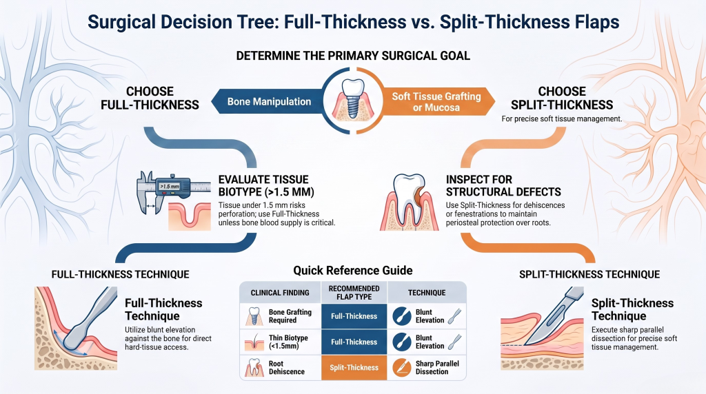
Decision Tree: Full-Thickness vs. Split-Thickness Flaps
Use this step-by-step logic when planning your next surgical case:
Assess the Surgical Goal:Determine if bone manipulation is required.
Determine if you need to alter, remove, or graft directly onto hard tissue. If you need direct visual access to bone, choose a Full-Thickness Flap. If your goal is soft tissue grafting or increasing attached mucosa, proceed to Step 2.
Evaluate Biotype and Bone Thickness:Check CBCT and clinical tissue thickness.
Measure the soft tissue biotype and inspect buccal bone thickness on your CBCT. If tissue is under 1.5 mm thick, sharp splitting carries a high risk of perforation. Consider a full-thickness elevation unless preserving bone blood supply is critical.
Inspect for Structural Defects:Look for fenestrations or dehiscences.
Check for dehiscences or fenestrations. If bare root surfaces exist under thin soft tissue, select a Split-Thickness Flap to leave periosteal protection over the defect.
Select Flap Design and Execute:Prepare dedicated instruments.
Select your incision design and blade type. Use blunt elevation against bone for full-thickness access, or sharp parallel dissection for split-thickness soft tissue management.
Post-Operative Management & Enhancing Team Outcomes
Clean surgical execution must be backed by consistent post-operative care:
Surgical Site Care Protocol: Instruct patients to avoid brushing the surgical site for 10 to 14 days. Prescribe a 0.12% Chlorhexidine gluconate rinse or antimicrobial spray to keep the area clean.
Interprofessional Team Roles: Train your dental assistants and hygienists to recognize normal healing versus early flap dehiscence, excessive swelling, or tissue pallor during follow-up visits.
Master Advanced Surgical Techniques with One World Dental
Reading surgical protocols is a great start, but true confidence comes from hands-on clinical practice. If you want to refine your surgical tissue handling, master soft tissue grafting, or perform complex implant procedures with ease, practical training is essential.
One World Dental offers comprehensive continuing education courses designed for general dentists and specialists looking to elevate their practice. Founded by Dr. Kambez Shukoor—a veteran and experienced clinician—One World Dental combines online learning with live, hands-on surgical training.
Why Train with One World Dental?
Tiered Learning Paths: Progress naturally from foundational soft tissue techniques to advanced surgical grafting and full-arch implant workflows.
Real-World Experience: Learn practical protocols you can apply in your practice immediately.
Mentorship-Driven Instruction: Receive real-time guidance from practicing clinicians who perform these surgeries daily.
Flexible Learning Formats: Access on-demand online lectures or participate in immersive hands-on workshops.
Whether you want to refine your flap reflection techniques, master connective tissue grafting, or expand your implant practice, One World Dental provides the step-by-step training you need.
People Also Ask (FAQs)
What is the main difference between a full-thickness and split-thickness flap?
A full-thickness flap reflects the mucosa, submucosa, and periosteum to expose bare bone. A split-thickness flap sharp-dissects through the tissue, lifting only the mucosa and part of the connective tissue while leaving the periosteum attached to the bone.
Does a full-thickness flap cause permanent bone loss?
Full-thickness reflection causes a small amount of temporary bone ischemia, typically resulting in about 0.5 mm to 1.0 mm of marginal bone resorption during early healing. In healthy patients with thick bone, this is generally negligible, but it can be significant in thin biotypes.
Why use a split-thickness flap for soft tissue grafting?
Leaving the periosteum attached to the bone preserves a highly vascular bed. When you place a connective tissue or free gingival graft on top of intact periosteum, the underlying blood supply nourishes the graft during early healing.
How thick must tissue be to safely perform a split-thickness flap?
Clinicians generally need at least 1.5 mm to 2.0 mm of tissue thickness to execute a split-thickness flap safely. Splitting tissue thinner than 1.5 mm increases the risk of tissue tears and partial flap necrosis.
What instruments are best for reflecting a split-thickness flap?
Split-thickness flaps require micro-surgical scalpel blades (such as a #15c, #64, or #69 blade) and fine micro-tissue forceps. Unlike full-thickness flaps, which use blunt elevators, split-thickness flaps rely on precise, sharp dissection.
Can general dentists learn these flap techniques through continuing education?
Yes. With structured clinical training, general dentists can master both full-thickness and split-thickness flap techniques. Comprehensive hands-on courses—like those offered by One World Dental—provide the guided practice needed to perform these procedures predictably.
Elevate Your Surgical Confidence Today
Mastering soft tissue management is the foundation of predictable implant dentistry, periodontics, and bone grafting. Don’t let uncertainty hold back your practice growth.
Educational disclaimer: Content is provided for professional education and is not a substitute for a clinician's independent judgment, applicable standards, manufacturer instructions, or relevant laws and regulations.
Practical chairside flow for flap design in implantology that protects the papilla. Includes a printable four point preop checklist and hands on training...