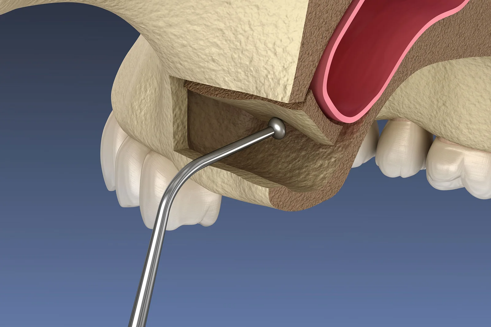
Level 2 • Implants & Surgery
Sinus Lift (Crestal & Lateral) – Online Lecture Series
Online lecture series on crestal and lateral window sinus augmentation approaches.
- Self-Paced
- 3–4 CE Credits
- Self-Paced
- On-Demand
Coming SoonView Course

Bone Grafting & Site Development • Comparison
Compare Crestal vs Lateral Window sinus lifts. Explore RBH thresholds, CBCT diagnostics, complications, and clinical criteria.
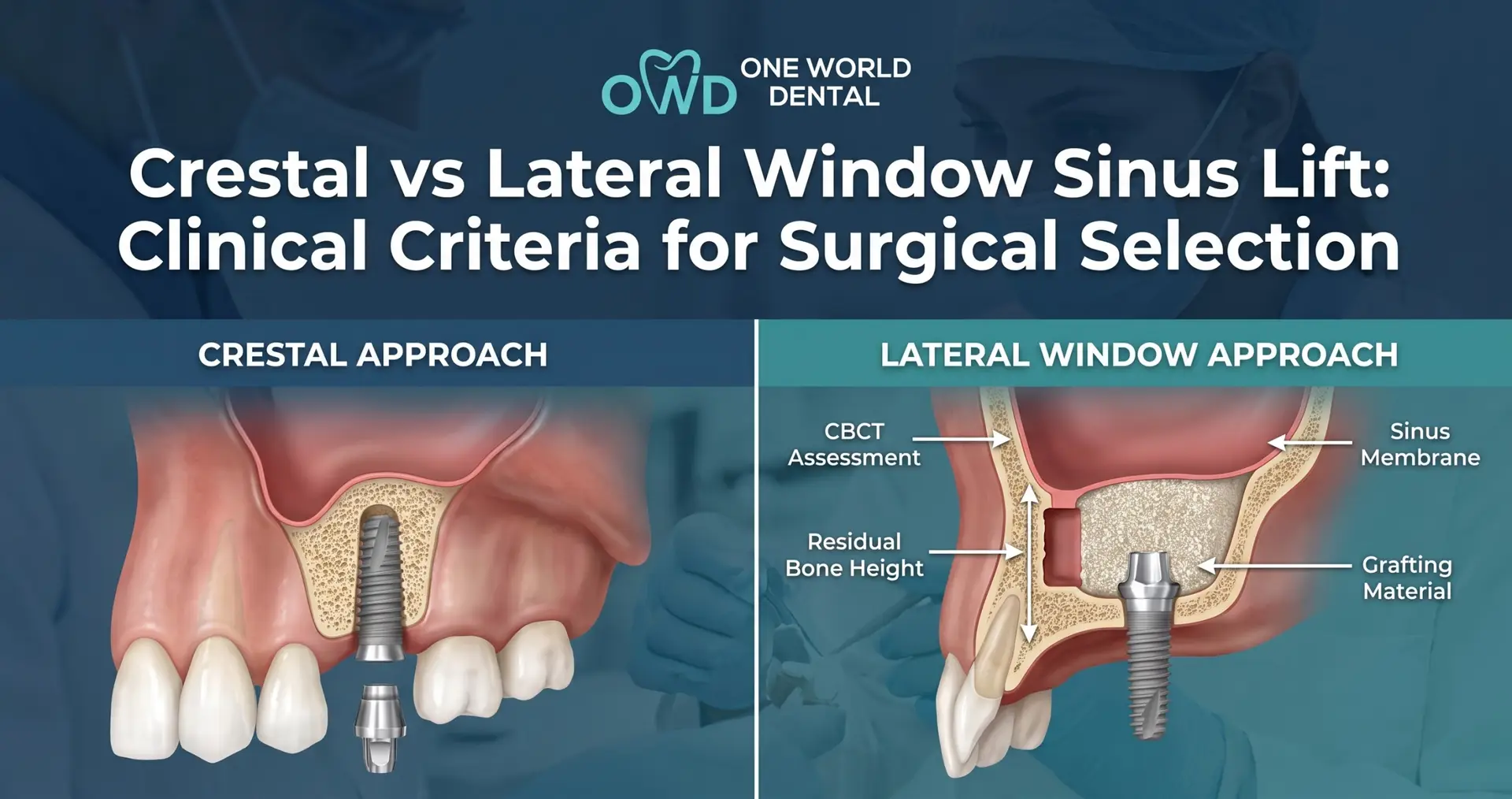
When selecting a surgical protocol for maxillary sinus floor elevation (MSFE), clinicians evaluating Crestal vs Lateral Window Sinus Lift protocols must balance surgical invasiveness against predictable implant stability. Reconstructing the atrophic posterior maxilla requires strict clinical criteria to choose between a transcrestal approach and a direct lateral access window. This evidence-based guide breaks down the anatomical thresholds, surgical workflows, and risk management strategies required to choose the right technique with total predictability.
The edentulous posterior maxilla presents unique anatomical challenges. Ridge resorption following tooth extraction, combined with pneumatization of the maxillary sinus, often leaves insufficient bone height for standard implant placement.
Maxillary sinus floor elevation (MSFE) restores the vertical dimension necessary to anchor endosseous implants. Since Hilt Tatum introduced the lateral window protocol and Robert Summers popularized the transcrestal osteotome technique, clinicians have debated where indirect elevation ends and direct surgical access becomes necessary. Choosing the correct approach directly impacts primary stability, operative morbidity, and overall treatment success.
Accurate treatment planning starts with high-resolution Cone-Beam Computed Tomography (CBCT). Traditional two-dimensional panoramic radiographs obscure crucial three-dimensional spatial relationships, leading to misdiagnoses.
+-------------------------------------------------------------------+
|                   KEY CBCT EVALUATION METRICS                      |
+-----------------------------------+-------------------------------+
| Anatomical Factor                 | Clinical Impact               |
+-----------------------------------+-------------------------------+
| Sinus Floor Width & Angle         | Narrow V-shape (<30°) increases|
|                                   | risk of membrane tearing.     |
| Presence of Underwood’s Septa     | Requires modified window design|
|                                   | or dual-window access.        |
| Membrane Thickness (1–2 mm ideal) | <0.5 mm tears easily;         |
|                                   | >3 mm indicates pathology.    |
| Lateral Sinus Wall Thickness      | Thick walls (>2 mm) favor     |
|                                   | piezoelectric osteotomy.      |
| Path of the PSAA                  | Intraosseous vessel must be   |
| (Posterior Superior Alveolar)     | mapped to avoid hemorrhage.   |
+-----------------------------------+-------------------------------+
Residual Bone Height (RBH) remains the primary diagnostic metric when choosing between crestal and lateral approaches.
     RESIDUAL BONE HEIGHT (RBH) SELECTION THRESHOLDS

  RBH ≥ 6–8 mm           RBH 4–6 mm           RBH < 4 mm
+--------------+       +------------+       +------------+
|   CRESTAL    |       | GREY ZONE  |       |  LATERAL   |
|   APPROACH   |       |  Indirect  |       |  WINDOW    |
|  (Predictable|       |     vs.    |       |  (Direct   |
|  Indirect)   |       |  Direct    |       | Visual)    |
+--------------+       +------------+       +------------+
The crestal technique accesses the sinus floor through the osteotomy created for implant placement.
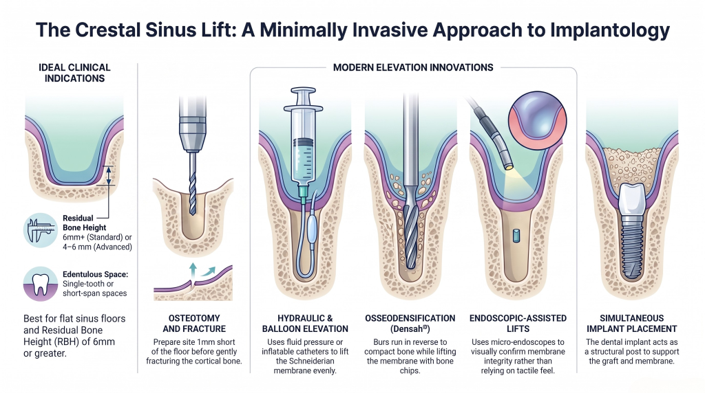
Perform osteotomy preparation up to 1 mm short of the sinus floor.
Gently fracture the cortical sinus floor using osteotomes, densifying burs, or hydraulic access drills.
Elevate the Schneiderian membrane using fluid pressure, specialized instruments, or a autogenous/xenograft bone pack.
Place the dental implant simultaneously to act as a structural post supporting the graft and membrane.
The lateral window technique creates a surgical access portal in the facial wall of the maxillary sinus, giving direct visual access to the Schneiderian membrane.
Elevate a full-thickness mucoperiosteal flap beyond the mucogingival junction.
Create an oval or rectangular osteotomy through the lateral maxillary wall using burs or piezoelectric tips.
Carefully detach and elevate the Schneiderian membrane from the sinus floor and internal walls using specialized curettes.
Pack the sub-antral cavity with selected biomaterials and place a resorbable barrier membrane over the lateral access window before suturing.
Choosing the optimal technique requires comparing clinical trade-offs across key performance metrics:
+---------------------------------------------------------------------------------------+
|                       CRESTAL VS. LATERAL WINDOW COMPARISON                           |
+-----------------------------------+-------------------------+-------------------------+
| Feature / Metric                  | Crestal (Indirect)      | Lateral Window (Direct) |
+-----------------------------------+-------------------------+-------------------------+
| Direct Membrane Visibility        | No (Blind technique)    | Yes (Full visual)       |
| Average Vertical Bone Gain        | 2–5 mm                  | 6–10+ mm                |
| Membrane Perforation Risk         | Moderate (Unseen)       | Higher total, but seen  |
| Primary Stability Potential       | High (requires RBH ≥5mm)| Low if RBH < 4 mm       |
| Surgical Morbidity / Swelling     | Low                     | Moderate to High        |
| Operative Time                    | Short (15–30 mins)      | Long (45–90 mins)       |
+-----------------------------------+-------------------------+-------------------------+
Selecting the right biomaterial ensures long-term graft stability and healthy bone turnover beneath the elevated membrane.
                BIOMATERIAL COMBINATION PROTOCOL

    +-------------------+           +-------------------+
    |   XENOGRAFT/      |           |    AUTOGRAFT/     |
    |   ALLOGRAFT       |   +---+   |    ALLOGRAFT      |
    |  Slow-Resorbing   | + |   | = | Fast-Turnover     |
    | Scaffold (70–80%) |   +---+   |   Osteogenic      |
    +-------------------+           |   Element (20–30%)|
                                    +-------------------+
                 MEMBRANE PERFORATION MANAGEMENT

                     Perforation Detected
                               |
            +------------------+------------------+
            |                                     |
     Crestal Approach                      Lateral Approach
            |                                     |
    +-------+-------+                     +-------+-------+
    |               |                     |               |
Small Tear      Large Tear            Small Tear      Large Tear
  (<3mm)          (>3mm)                (<5mm)          (>5mm)
    |               |                     |               |
Place PRF/      Abort &               Cover with      Suture /
Collagen;       Re-evaluate           Resorbable      Matrix Slate /
Pack Gently     in 3–4 mos.           Membrane        Abort Site
If an osteotomy cuts the PSAA during a lateral lift, apply direct pressure using a gauze soaked in epinephrine, use electrocautery, or crush the surrounding bony canal with a dull instrument to achieve hemostasis before continuing.
Bacterial contamination can cause acute graft infection or maxillary sinusitis. Manage early signs using systemic broad-spectrum antibiotics (such as Amoxicillin/Clavulanate), nasal decongestants, and warm saline rinses. Unresolved graft infections require surgical drainage and curettage.
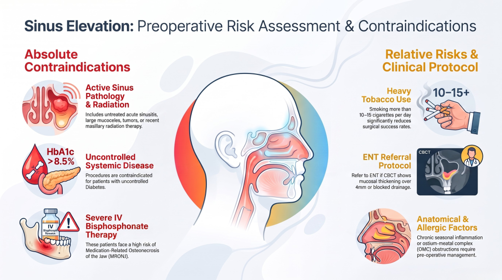
Clinical Protocol: When pre-operative CBCT scans show mucosal thickening over 4 mm or a blocked sinus ostium, refer the patient to an ENT specialist to establish normal drainage before attempting sinus elevation.
Follow this systematic decision tree to select the safest, most predictable surgical workflow:
                  CBCT DIAGNOSTIC SELECTION WORKFLOW
                                  |
                      Evaluate RBH at Site
                                  |
        +-------------------------+-------------------------+
        |                                                   |
   RBH ≥ 6–8 mm                                        RBH < 4 mm
        |                                                   |
Check Sinus Floor Morphology                         Select LATERAL WINDOW
        |                                                   |
 +------+------+                                     +------+------+
 |             |                                     |             |
Flat         Steep/Septa                           RBH < 3mm    RBH 3–4mm
 |             |                                     |             |
Select       Select                                Staged       Simultaneous
CRESTAL      LATERAL                               Placement    Placement
APPROACH     WINDOW                                (Wait 6-9m)  (If stable)
Measure Residual Bone Height (RBH) on CBCT.
If RBH is in the 4 to 6 mm grey zone, evaluate local anatomy:
Evaluate Primary Stability Expectations:
Mastering advanced sinus elevation techniques requires more than reading clinical literature—it demands hands-on experience, expert instruction, and tactical feedback. Transitioning confidently from conservative crestal lifts to complex lateral window surgeries is a major milestone for growing dental practices.
At One World Dental, we bridge the gap between classroom theory and clinical execution. Led by experienced surgical educators, our evidence-based continuing education platform offers comprehensive training tailored to your clinical skill level.
Whether you want to refine your crestal techniques or master lateral window procedures with hands-on live-patient mentorship, One World Dental provides the practical skills you need to transform your practice.
Take the next step in elevating your surgical practice today. Contact One World Dental to view our course catalog and register for upcoming surgical training programs.
A traditional osteotome crestal sinus lift usually requires at least 5 to 6 mm of native residual bone height (RBH) to ensure primary implant stability. However, using modern techniques like osseodensification burs or hydraulic elevation systems, experienced clinicians can predictably execute crestal lifts with as little as 4 mm of native bone.
The most common cause of intraoperative sinus lift failure is Schneiderian membrane perforation that goes unrecognized or unmanaged. Unrepaired tears allow bone graft material to migrate into the sinus cavity, leading to acute or chronic sinusitis, graft infection, and loss of both the graft and the dental implant.
Yes. Studies show that gently elevating the Schneiderian membrane and placing an implant simultaneously creates a protected space beneath the membrane. The resulting blood clot can naturally organize and form new bone around the implant apex without needing added particulate bone grafts, provided the membrane remains intact.
If you suspect an indirect perforation (evidenced by a loss of tactile resistance or lack of membrane rebound), do not force particulate bone into the osteotomy. Instead, place a resorbable collagen sponge or a PRF plug into the top of the osteotomy to seal the area. If the tear seems large, do not elevate further; place the implant short of the sinus floor or abort and allow the site to heal for 3 to 4 months.
Implants placed simultaneously with a sinus lift typically require a healing period of 4 to 6 months to allow the surrounding graft material and native bone to integrate fully. If the native bone height was severely compromised (under 4 mm) or initial insertion torque was low, allow 6 to 9 months of unweighted healing before prosthetic loading.
Piezosurgery uses ultrasonic micro-vibrations tailored specifically to cut mineralized bone while sparing soft tissue. If the vibrating tip touches the delicate Schneiderian membrane, it does not tear it. This significantly lowers perforation rates compared to traditional rotary carbide or diamond burs.
You can identify the location, path, and diameter of the intraosseous PSAA using pre-operative CBCT cross-sectional views. Knowing the exact distance from the alveolar crest to the artery allows you to position your upper horizontal osteotomy cut above or below the vessel, preventing intraoperative arterial bleeding.
Educational disclaimer: Content is provided for professional education and is not a substitute for a clinician's independent judgment, applicable standards, manufacturer instructions, or relevant laws and regulations.
Related Training
Level 2 • Implants & Surgery
Online lecture series on crestal and lateral window sinus augmentation approaches.
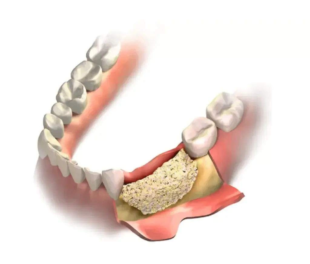
Level 1 • Implants & Surgery
Self-paced online lecture series on bone graft materials, membranes and biologics: how they work, how to choose them and how to handle them.
Bone Grafting • Clinical Guide
Master the lateral sinus lift technique to enhance bone volume for implants. Learn when to choose this method over others for optimal results.
23 min read · Updated July 27, 2026
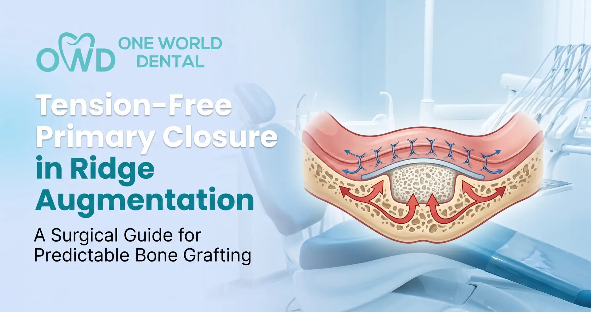
Bone Grafting • Procedure Guide
Master step-by-step flap release, suturing, and soft tissue management to achieve tension-free primary closure and prevent graft dehiscence.
10 min read · Updated August 11, 2026
Bone Grafting • Professional Education
Master clinical risk assessment for ARP. Learn to identify systemic, anatomical, and intra-operative red flags that necessitate specialist.
7 min read · Updated May 22, 2026