Distinguishing between FP1 vs. FP2/FP3 restorations represents a foundational diagnostic threshold in full-arch implant prosthodontics. Misjudging the prosthetic boundaries during treatment planning introduces significant risks: catastrophic biomechanical failure, phonetic distortion, irreversible aesthetic compromise, or non-cleansable contours that accelerate peri-implantitis. Executing a predictable full-arch rehabilitation demands strict adherence to objective anatomical, spatial, and mechanical parameters.
Introduction to Carl Misch’s Fixed Prosthesis (FP) Classification
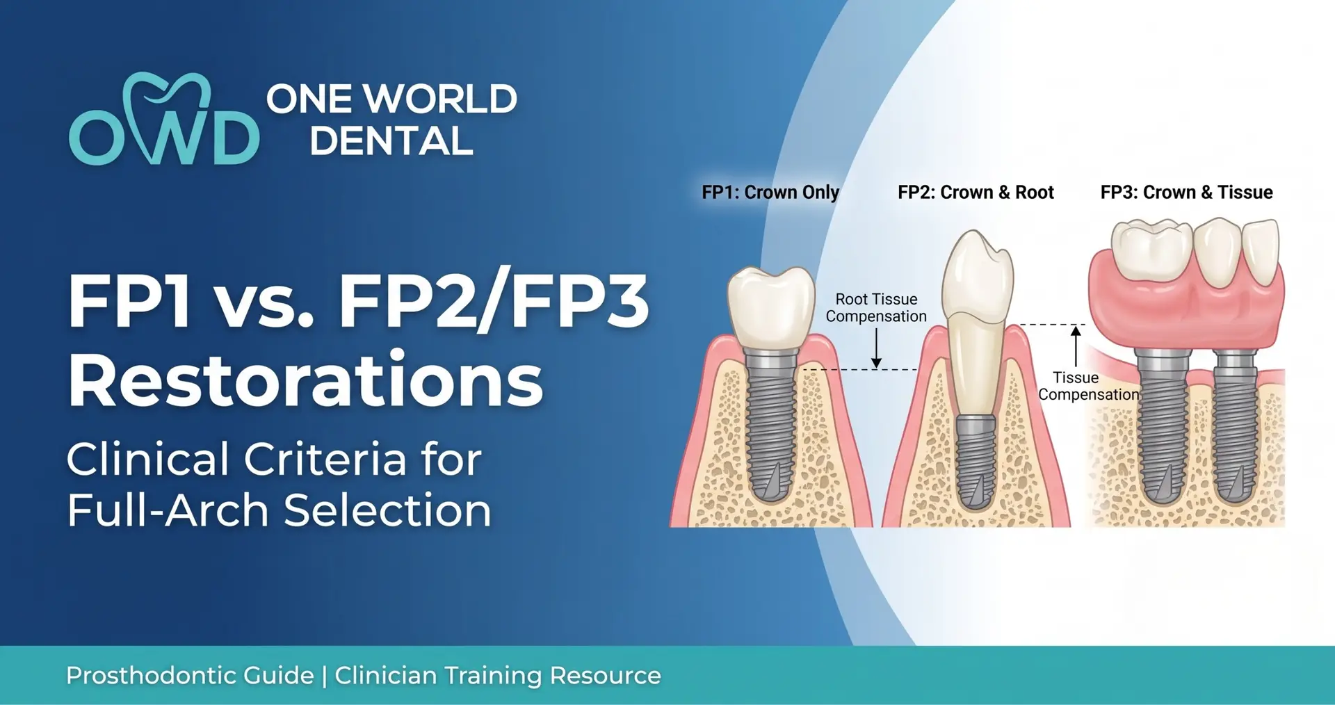
Dr. Carl Misch’s foundational classification system categorizes implant prostheses based on the specific anatomical structures they replace, establishing a direct relationship between bone resorption and prosthetic design.
The Anatomical Crown Replacement An FP1 restoration replaces only the missing clinical crowns. The emergence profile mimics natural dentition, with the prosthetic margins terminating within a healthy, intact peri-implant mucosal architecture.
The Hypercontoured, Elongated Root Solution An FP2 restoration replaces both the clinical crown and the cervical portion of the root structure. The clinical crowns appear elongated, and the design is indicated only when the junction is fully masked by the patient’s lip dynamics.
The “Pink & White” Tissue-Compensated Hybrid An FP3 restoration replaces the clinical crowns as well as lost alveolar ridge volume. It utilizes pink porcelain, layered composite, or acrylic to mask the underlying ridge deficiency and restore proper lip support.
Glossary of Critical Prosthodontic Terminology
Alveolar Ridge Resorption: The multi-dimensional centripetal and vertical bone loss following tooth loss (Seibert/Misch Classifications).
Emergence Profile: The axial contour of a prosthesis extending from the implant platform or multi-unit abutment (MUA) through the peri-implant mucosa to the visible clinical environment.
Transition Line: The prosthetic-mucosal junction where the artificial gingiva of an FP3 prosthesis meets the patient’s residual ridge.
The Multiphase Diagnostic Protocol
Clinical Visual Assessment & Diagnostic Wax-Ups Initial assessment must evaluate vertical dimension of occlusion (VDO), maxillomandibular relationships, and phonetic baselines sounds. A comprehensive diagnostic wax-up or virtual tooth arrangement sets the baseline for the restorative envelope.
CBCT Bone Volume and Density Analysis (Misch Bone Divisions) Cone-Beam Computed Tomography (CBCT) cross-sectional imaging maps available bone volume according to Misch Divisions (A through D). Division A allows for primary FP1 placement, whereas Division C/D requires substantial augmentation or an FP3 pathway.
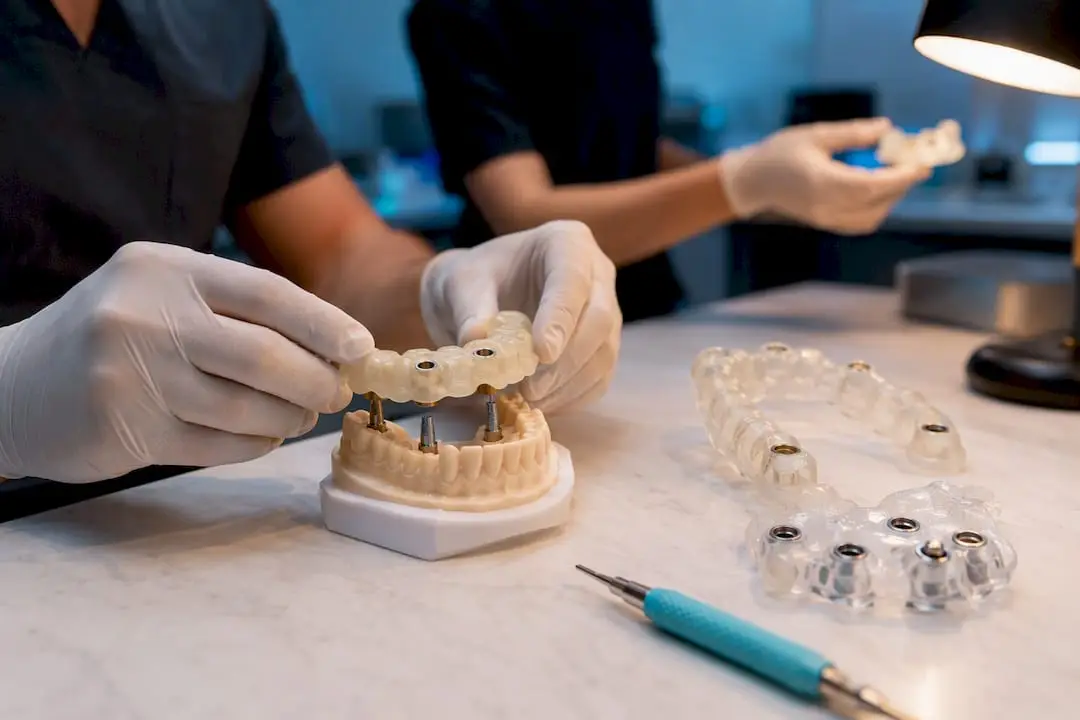
Verifying Three-Dimensional Implant Positions and Surgical Guides Prosthetically-driven implant placement utilizes stackable, digitally designed surgical templates. These guides ensure that the implant fixtures are positioned relative to the planned emergence profile rather than merely following residual bone morphometry.
Core Clinical Criteria for Selection: The Decision Matrix
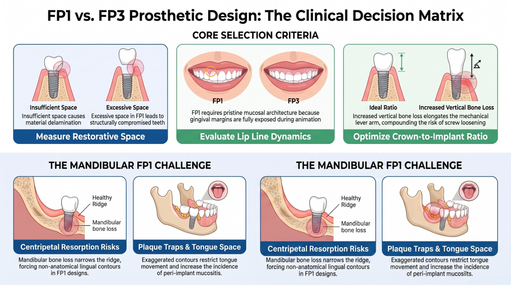
Available Restorative Space (Vertical and Horizontal Measurements) Restorative space represents the distance from the implant platform/MUA to the planned incisal edge or occlusal plane. Insufficient space causes material delamination or framework fracture; excessive space in an FP1 configuration creates structurally compromised, hyper contoured teeth.
Lip Lines and Dynamics:The Maxillary Smile Line vs. Mandibular Sibilant Sounds A high or hypermobile maxillary lip line demands that the transition line of an FP3 restoration sits coronal to the smile line, or far apical to it. For an FP1, the mucosal architecture must be pristine, as the gingival margins are completely exposed during animation.
Crown-to-Implant Ratio and Biomechanical Force Distribution As vertical bone loss increases, the mechanical lever arm elongates. An unfavorable crown-to-implant ratio increases micro-strain at the crestal bone interface, compounding the risks of screw loosening, component fatigue, or marginal bone loss under lateral loads.
Maxillary vs. Mandibular Design Realities: Why Mandibular FP1s Carry Higher Risk Executing a true mandibular FP1 is exceptionally challenging due to the pattern of mandibular resorption, which proceeds centripetally and inferiorly, narrowing the basal ridge. Attempting an FP1 design on a resorbed mandible results in exaggerated, non-anatomical lingual contours and restricted tongue space. These cantilevers act as immediate plaque traps, drastically increasing the incidence of peri-implant mucositis.
The Diagnostic “Math”: Restorative Space Requirements by Case Type
Prosthetic selection must be governed by precise linear measurements from the restorative platform to the planned incisal edge or occlusal plane.
Restoration Classification
Minimum Required Vertical Interarch Space
FP1 (Single/Splinted)
$8 – 10 \text{ mm}$
FP2 (Elongated Roots)
$10 – 12 \text{ mm}$
FP3 (Monolithic Zirconia / Titanium-Bar)
$12 – 15+ \text{ mm}$
FP1 Spatial Window: The 8–10 mm Constraint for Natural Emergence An FP1 restoration requires a strict, constrained vertical envelope of 8 to 10 mm. Exceeding 10 mm of space results in unacceptably long clinical crowns (FP2 territory) or requires a soft tissue grafting protocol to coronalize the peri-implant mucosa.
FP2 Spatial Window: Managing Intermediate Moderate Loss Operating within the 10 to 12 mm window, the FP2 design compensates for moderate vertical deficiency by extending the cervical contours. This approach relies on a low lip line to conceal the exposed pseudo-roots.
FP3 Spatial Window: The 12–15+ mm Minimum for Zirconia and Titanium-Acrylic Hybrids An FP3 configuration requires a minimum of 12 to 15 mm of space. This depth accommodates the substructure framework (titanium bar or zirconia core), the overlay material (acrylic, composite, or porcelain), and the necessary multi-unit abutment heights while preserving material structural integrity.
Scalloped Tissue Architecture vs. Flat Ridge Recontouring FP1 designs demand a highly managed, scalloped peri-implant mucosal architecture mimicking natural interdental papillae. Conversely, FP3 designs require aggressive surgical alveoloplasty to flatten the residual ridge, ensuring a smooth, non-retentive prosthetic interface.
Soft Tissue Augmentation: Achieving Natural Emergence Profiles in FP1 Restorations To manage the transition zones of an FP1 full-arch prosthesis, advanced soft tissue augmentation—including subepithelial connective tissue grafts (SCTG) or free gingival grafts (FGG)—is mandatory to restore lost horizontal and vertical tissue volume, ensuring a stable, cleansable sulcus.
The Role of Fixed Provisionals in Guiding Tissue Healing Polymethyl methacrylate (PMMA) or bis-acrylic provisional restorations are critical for dynamic tissue sculpting. Over weeks of healing, the emergence profiles are modified via additive or subtractive adjustments to guide the peri-implant mucosa into the desired scalloped form.
Biomechanical Engineering & Dental Material Selection
Milled Monolithic Zirconia vs. PFM Substructures Milled monolithic zirconia provides superior flexural strength ($>1000 \text{ MPa}$) and excellent biocompatibility, making it an ideal choice for high-load FP3 arches. PFM configurations provide precise, isolated margins suitable for multi-unit FP1 applications.
Milled Titanium Frameworks Overlaid with Acrylic/Composite This traditional choice utilizes a rigid, CAD/CAM-milled titanium substructure wrapped in PMMA or composite. This assembly provides high impact energy absorption, reducing peak loads transmitted to the underlying bone interface.
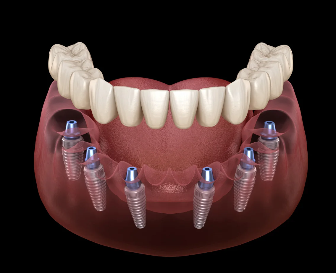
Implant Numbers and Surface Area: 4 Implants vs. 6-8+ Anchorage Requirements While an FP3 design can often be executed via an All-on-4 configuration by tilting distal fixtures, an FP1 architecture lacks pink acrylic to mask long cantilevers. FP1 cases typically require 6 to 8 implants to limit distal cantilevers and provide adequate support along a natural arch form.
Occlusal Force Management: Frankfurt Mandibular Angle (FMA) Impact on Fracture Risk Patients exhibiting a low FMA (brachyfacial/hyperdivergent skeletal patterns) possess masseteric hypertrophy, generating elevated nocturnal parafunctional forces. These heavy biters pose a severe threat to monolithic zirconia or porcelain overlays, demanding rigid metal-backed substructures, strict mutually protected occlusal schemes, and mandatory nocturnal bruxism appliances.
Retention Strategies: Screw-Retained vs. Cement-Retained Systems Screw retention remains the gold standard for full-arch fixed prostheses, ensuring retrievability and eliminating the risk of subgingival cement sepsis. Cement-retained solutions are generally restricted to FP1 cases where the implant axis emerges through the incisal edges of anterior teeth, though angled screw channels (ASC) have largely superseded this approach.
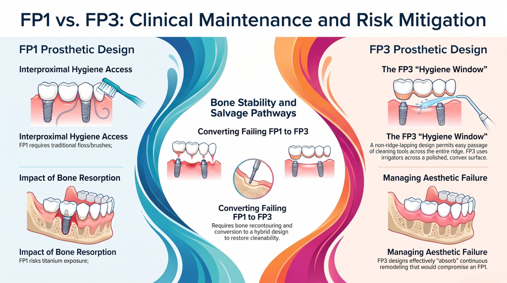
The Hygiene Window: Designing Cleanable Interproximal Spaces for FP1 vs. FP3 An FP1 requires meticulous interproximal hygiene access via traditional interdental brushes and floss. An FP3, conversely, must feature a highly polished, convex, non-ridge-lapping tissue surface. This design permits a clear hygiene window for the passage of oral irrigators and proxy-brushes across the entire ridge.
Dealing with Bone Resorption and Gum Recession Over Time Late-stage crestal bone loss compromises the marginal integrity of an FP1, exposing the titanium implant collars or abutment junctions. In contrast, an FP3 design effectively absorbs this continuous remodeling, since its prosthetic-tissue junction remains hidden beneath the lip line.
Reversible Pathways: Converting a Failing FP1 to an FP3 Masterpiece When chronic peri-implantitis or aesthetic failure occurs in an FP1 system, clinicians must plan a salvage pathway. This typically requires removing the prosthesis, performing targeted bone recontouring or guided bone regeneration (GBR), and converting the prosthetic design into an FP3 hybrid to re-establish proper biomechanics and cleanability.
Conclusion: Achieving Predictable Results and Patient Satisfaction
Summary Matrix for Clinical Decision-Making Selecting the appropriate fixed prosthesis classification is a highly technical workflow that balances anatomical limitations against structural material requirements. Practitioners must measure vertical space, map lip dynamics, and analyze skeletal load profiles to ensure long-term clinical success.
References & Evidence-Based Literature Review Long-term clinical data underscores that forcing an FP1 profile onto an inadequate hard- and soft-tissue foundation introduces a significant risk of prosthetic failure and peri-implant bone loss. Pre-operative digital planning remains indispensable for identifying these boundaries.
When managing complex full-arch cases that require high-precision CAD/CAM milling and strict adherence to prosthodontic principles, partner with a technical team that understands these exact criteria. Visit One World Dental to consult on your practice’s next advanced multi-unit case evaluation and diagnostic wax-up.
What is the primary indicator for converting an intended FP1 full-arch case into an FP2 or FP3 design mid-treatment? The primary indicator is an insufficient vertical mucosal thickness or severe multi-directional ridge resorption observed during the diagnostic wax-up. If the vertical distance from the implant platform to the planned incisal edge exceeds $10 \text{ mm}$, attempting an FP1 will yield non-anatomical, hypercontoured crown profiles that create unmanageable plaque traps.
How does the choice between FP1 and FP3 impact the selection of multi-unit abutment (MUA) heights? FP1 restorations require very short MUAs ($1 – 2 \text{ mm}$) or direct-to-fixture configurations to ensure that the prosthetic margin remains subgingival or at the mucosal crest. FP3 restorations require taller MUAs ($3 – 5 \text{ mm}$) to project the restorative platform above the recontoured tissue level, ensuring a highly cleansable, convex interface.
Why does an FP1 configuration frequently necessitate a greater number of implants compared to an All-on-4 FP3 design? An FP1 design follows a natural, scalloped arch anatomy without the structural reinforcement of a thick, continuous acrylic or zirconia base. To prevent dangerous tensile and shear forces on cantilevers, the anterior-posterior (A-P) spread must be optimized by distributing the functional load across 6 to 8 implants.
How do you manage phonetic issues when transitioning from a temporary provisional to a final FP3 prosthesis? Phonetic issues with an FP3 usually stem from air escaping through an improper tissue-prosthesis interface or from excessive palatal bulk. The provisional must be modified with flowable composite to optimize the seal against the ridge. Once phonetics are verified, the contour must be duplicated precisely via a double-scan technique for the final milled prosthesis.
Is monolithic zirconia contraindicated for an FP1 full-arch prosthesis in patients presenting with severe bruxism? While not completely contraindicated, it presents a very high clinical risk. An FP1 lacks the bulk of an FP3 hybrid, making isolated interproximal connectors susceptible to fatigue fracture under parafunctional loads. If monolithic zirconia is selected, the connectors must have a minimum cross-sectional area of $9 – 12 \text{ mm}^2$, and a heavy-duty nocturnal occlusal splint is mandatory.
What specific surgical step is mandatory during the placement of an FP3 restoration that is avoided in an FP1 protocol? An FP3 restoration requires a strategic, aggressive alveoloplasty (bone reduction) during surgery to create at least $12 – 15$ of vertical restorative space and hide the transition line. In contrast, an FP1 protocol prioritizes preserving crestal bone height and often requires bone or soft-tissue grafting to maintain the natural alveolar ridge profile.
Educational disclaimer: Content is provided for professional education and is not a substitute for a clinician's independent judgment, applicable standards, manufacturer instructions, or relevant laws and regulations.
Prosthetic first digital workflow for clinicians: plan full arch implants, enable same day provisionals, target a 6 week definitive, and validate guides...