Avoiding the Collapse: A Definitive Guide to Managing the Thin Buccal Plate
Master advanced protocols for the thin buccal plate. Learn immediate implant placement, "Dual Zone" grafting, and tissue preservation.
Publisher
Published by One World Dental
Published
Published May 20, 2026
Reading time
6 min read
On this page
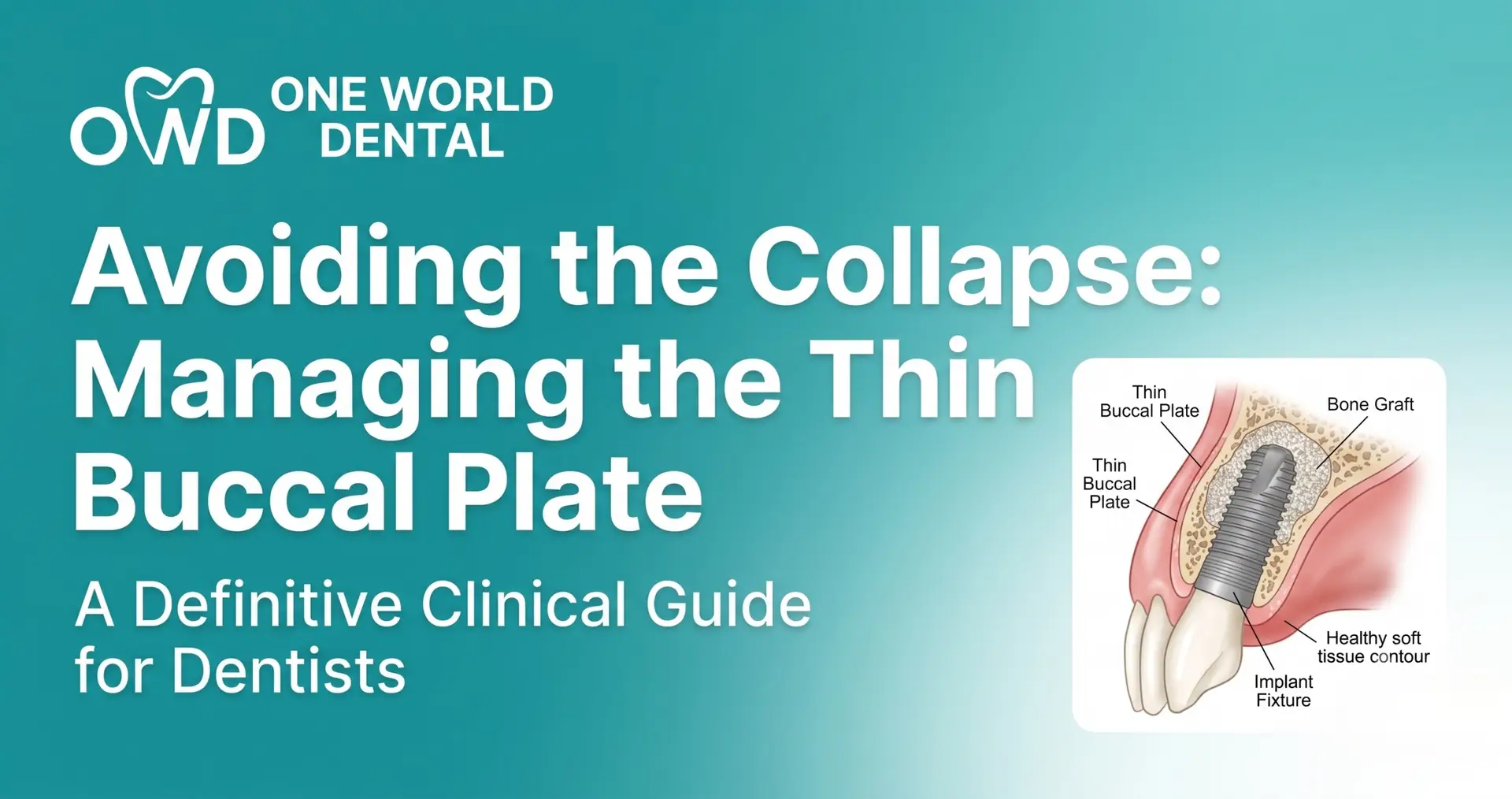
To achieve clinical success in the esthetic zone, mastering the management of the thin buccal plate is a foundational skill for the modern implantologist. Avoiding the collapse of the alveolar ridge requires a meticulous synergy between atraumatic extraction, three-dimensional implant positioning, and advanced grafting protocols. Through the evidence-based curriculum at One World Dental, practitioners can refine these techniques to ensure long-term hard and soft tissue stability.
The Biological Dilemma of the Thin Buccal Plate
The buccal plate in the anterior maxilla is often less than 1 mm thick. This bone is primarily “bundle bone”—a tooth-dependent structure that contains the Sharpey’s fibers of the periodontal ligament.
Anatomy of the “Bundle Bone” and its Vulnerability
Bundle bone is highly vascularized via the periodontal ligament. When a tooth is extracted, this primary blood supply is severed. Because the thin buccal plate lacks a significant cancellous marrow component, it is prone to rapid osteoclastic resorption.
Why the Buccal Plate Collapses Post-Extraction
Without the internal blood supply from the tooth, the thin cortical shell relies solely on the periosteum. If the periosteum is reflected (raised in a flap), the external blood supply is also compromised. This “double insult” leads to significant horizontal and vertical ridge contraction, often referred to as the “collapse.”
Defining the “Thin” Phenotype: Measurement and Classification
Practitioners must categorize the biotype before the first incision. A “thin” phenotype is clinically defined as a buccal plate of ≤1.0 mm. Using a periodontal probe, if the shadow of the probe is visible through the gingival margin, the patient possesses a thin soft tissue biotype, which almost always correlates with an underlying thin buccal plate.
Risk Assessment and Pre-Surgical Planning
Predictable outcomes start with advanced imaging and a conservative mindset.
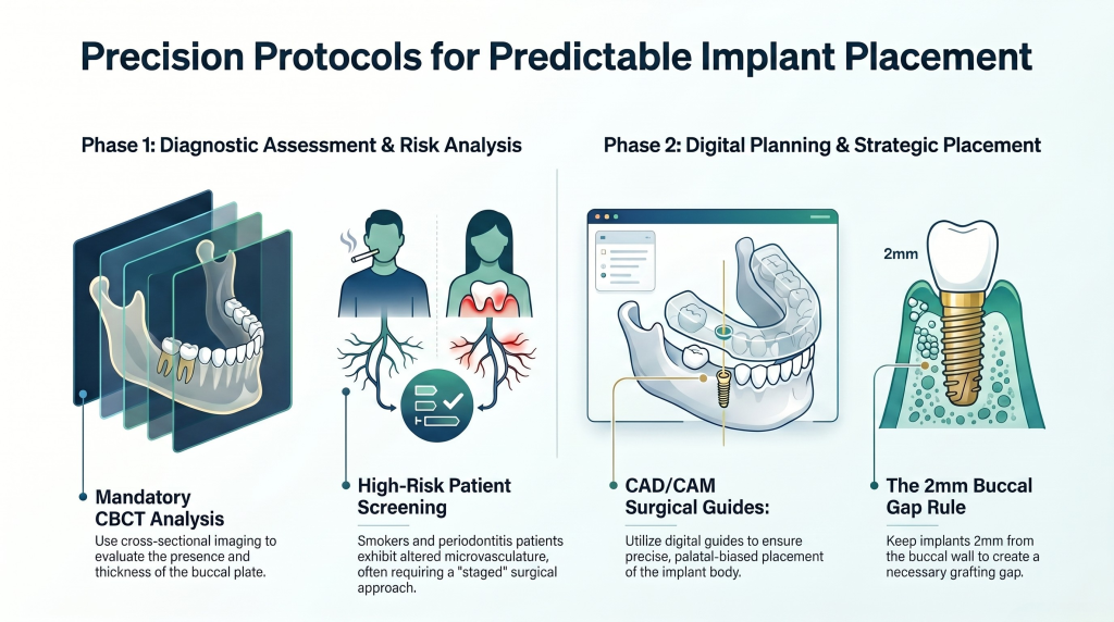
CBCT Analysis: Cross-sectional imaging is mandatory to evaluate the presence and thickness of the buccal plate.
Identifying High-Risk Patients: Smokers and patients with active periodontitis exhibit altered microvasculature. These factors accelerate bone resorption. At One World Dental, we emphasize that high-risk biotypes may require a “staged” approach rather than immediate placement.
Digital Planning: Utilizing CAD/CAM surgical guides allows for “palatal-biased” placement, keeping the implant body at least 2 mm away from the thin buccal wall to allow for a “gap” that can be grafted.
Surgical Protocols for Tissue Preservation
Flap vs. Flapless Approaches
A flapless approach is preferred for thin buccal plates. By keeping the periosteum attached to the bone, you preserve the external blood supply. If a flap is necessary for visibility, it should be a “split-thickness” flap where possible to maintain some vascularity to the bone.
Atraumatic Extraction Techniques
The use of periotomes and piezo-surgical inserts is critical. The goal is to sever the PDL without applying any pressure to the buccal plate. Forceps should only be used once the tooth is highly mobile.
The Socket-Shield Technique (Partial Extraction Therapy)
This technique involves leaving the buccal fraction of the tooth root in situ to maintain the attachment of the bundle bone.
Case Selection: Ideal for healthy, non-infected roots in the esthetic zone.
Common Pitfalls: Failure to decapitate the root 1mm below the bone crest or accidental displacement of the shield during implant osteotomy.
Immediate Implant Placement (IIP) Strategies
Management of the Buccal Gap: The “Dual Zone” Concept
The “Dual Zone” refers to the bone zone (the gap between the implant and the bone) and the tissue zone (the space created by the contour of the abutment). Even in tight gaps, “jumping distances” should be filled with a low-substitution bone graft to provide long-term volume.
Selecting the Correct Implant Diameter
A common error is using an implant that is too wide. In a thin buccal plate scenario, a narrower implant (3.3mm to 3.8mm) is often safer. This maintains a larger “gap” for grafting and prevents pressure necrosis of the cortical bone.
Palatal/Lingual Positioning
The implant should be positioned toward the palatal wall of the socket. This ensures that the entire diameter of the implant is supported by native bone while leaving the buccal aspect free for augmentation.
Advanced Grafting and Augmentation
Bone Grafting Materials
Xenografts: Bovine-derived bone (e.g., Bio-Oss) is the gold standard for thin plates because it resorbs very slowly, acting as a permanent scaffold.
Allografts: Useful for faster remodeling but may not provide the long-term volume stability needed to prevent collapse.
Contour Augmentation
Even if the plate is intact, “over-bulking” the buccal aspect with a slow-resorbing graft and a collagen membrane can compensate for the minor remodeling that will inevitably occur.
The Role of PRF in Soft Tissue Healing
Platelet-Rich Fibrin (PRF) contains growth factors that accelerate soft tissue closure. Placing PRF over a grafted socket-shield or immediate implant site protects the graft and improves the thickness of the overlying mucosa.
Soft Tissue Management: The Second Line of Defense
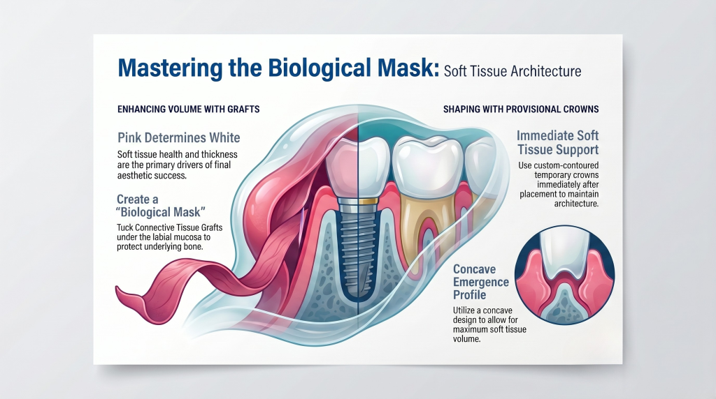
In many cases, the “pink” determines the “white.”
Connective Tissue Grafts (CTG): If the buccal plate is extremely thin, a CTG can be “tucked” under the labial mucosa. This increases the soft tissue thickness, which creates a “biological mask” and protects the underlying bone from the environment.
Provisional Crowns: A custom-contoured temporary crown should be used to support the soft tissue architecture immediately after placement. A concave emergence profile allows for maximum soft tissue volume.
Complications and Troubleshooting
Fenestration: If you feel the drill “give” or see a hole in the buccal plate, you must convert to a guided bone regeneration (GBR) procedure.
Gingival Recession: Often caused by an implant placed too far labially. Correction usually requires removing the implant or extensive soft tissue grafting.
Rescue Protocols: If the buccal plate fractures during extraction, abort immediate placement. Perform a “Socket Preservation” graft, wait 4-6 months, and return for a delayed implant.
Post-Operative Care and Long-Term Maintenance
Loading Protocols
For thin-plate cases, “non-functional” immediate loading is ideal. The provisional crown should be out of occlusion in both centric and excursive movements.
Patient Compliance
Oral hygiene is paramount. Biofilm accumulation on a thin-plate implant leads to rapid “perimucositis,” which can turn into “peri-implantitis” faster than in areas with thick cortical bone.
Conclusion: The Future of Ridge Preservation
Managing the thin buccal plate is a high-stakes endeavor that requires a deep understanding of biology and biomechanics. By utilizing the “Dual Zone” concept, atraumatic techniques, and proper material selection, practitioners can achieve flawless results.
One World Dental provides the advanced surgical training necessary to master these complex protocols. Elevate your clinical expertise and gain the confidence to handle the most challenging esthetic cases.
Can I place an implant if the buccal plate is completely missing? In cases of a missing buccal plate (Type 2 or 3 sockets), immediate placement is highly risky. Usually, a staged approach with Guided Bone Regeneration (GBR) is required to re-establish the bony wall before implant placement.
What is the “Jumping Distance” in implant dentistry? This is the gap between the implant surface and the internal wall of the extraction socket. If this gap is >2mm, it must be grafted to ensure the bone grows to the implant surface and maintains the ridge contour.
Why is a xenograft preferred over an allograft for ridge preservation? Xenografts (like bovine bone) have a very slow resorption rate. In the buccal plate area, we want a material that stays in place for years to provide structural support against the pressure of the lip and facial muscles.
Is the Socket-Shield technique considered standard of care? While it is supported by significant clinical literature, it is considered an advanced technique. It requires specialized training, such as that offered at One World Dental, to ensure the root fragment does not become infected or displaced.
How long should I wait to load an implant in a thin buccal plate? If primary stability is high (>35 Ncm), immediate non-functional loading can be performed. If stability is low, a submerged healing period of 3 to 4 months is recommended to allow for full osseointegration and graft maturation.
What happens if I use an implant that is too wide? A wide implant reduces the space for a bone graft and may put direct pressure on the thin cortical bone. This often leads to pressure necrosis, causing the bone to resorb and the gingiva to recede.
Educational disclaimer: Content is provided for professional education and is not a substitute for a clinician's independent judgment, applicable standards, manufacturer instructions, or relevant laws and regulations.
For clinicians: a defect-based ridge augmentation guide that matches technique to anatomy, explains mesh and CAD/CAM workflows, plans 4–6 month staging,...